Kevin Jiang, MD | Orthopaedic Surgery & Sports Medicine in Queens, NY
Kevin Jiang, MD
Orthopaedic Surgery · Sports MedicineCirugía Ortopédica · Medicina Deportiva骨科外科 · 运动医学
EN
EN
ES
ES
中文
中文
AboutSobre el Dr. Jiang关于姜医生
ConditionsLesiones y afecciones疾病与损伤
AthletesAtletas运动员
LocationsConsultorios诊所位置
SurgeryCirugía手术须知
Call (866) 670-6824Llamar (866) 670-6824致电 (866) 670-6824
Request an AppointmentSolicitar una cita预约就诊

Experienced care,Atención con experiencia,值得信赖的专科诊疗,
here in Queens.aquí en Queens.就在皇后区。

Kevin Jiang, MD is a board-certified orthopaedic surgeon with subspecialty certification in sports medicine at NewYork-Presbyterian Queens. Dr. Jiang specializes in nonoperative and operative treatment of sports injuries of the shoulder, elbow, and knee, and treats pediatric, adolescent, and adult patients. Dr. Jiang performs arthroscopic, minimally invasive, and complex open surgery, and has been an attending orthopaedic surgeon since 2014.Kevin Jiang, MD es cirujano ortopédico certificado por la junta, con certificación de subespecialidad en medicina deportiva en NewYork-Presbyterian Queens. Se especializa en el tratamiento quirúrgico y no quirúrgico de lesiones deportivas del hombro, el codo y la rodilla, y atiende a pacientes pediátricos, adolescentes y adultos. Realiza cirugía artroscópica, mínimamente invasiva y cirugía abierta compleja, y es cirujano ortopédico de planta desde 2014.Kevin Jiang 医学博士是获得美国骨科委员会认证的骨科外科医生,并具有运动医学专科认证,任职于 NewYork-Presbyterian Queens。姜医生专长于肩、肘、膝运动损伤的手术与非手术治疗,接诊儿童、青少年及成人患者,并开展关节镜手术、微创手术以及复杂的开放手术。自 2014 年起担任骨科主治医师。

With practice locations in Jackson Heights, Long Island City, and Flushing, he is committed to high-quality orthopaedic care in Queens. Our team will help diagnose your condition, understand the treatment options, and help get you back to your activities.Con consultorios en Jackson Heights, Long Island City y Flushing, está comprometido con una atención ortopédica de alta calidad en Queens. Nuestro equipo le ayudará a diagnosticar su afección, a entender las opciones de tratamiento y a volver a sus actividades.诊所设于 Jackson Heights、Long Island City 与 Flushing,致力于为皇后区提供高质量的骨科诊疗。我们的团队将帮助您明确诊断、了解治疗方案,并协助您重返日常活动。

Dr. Kevin Jiang, MD, in a NewYork-Presbyterian Queens white coat, standing in front of his framed diplomas.
Assistant Professor of Clinical Orthopedic SurgeryProfesor Asistente de Cirugía Ortopédica Clínica临床骨科外科助理教授
Weill Cornell Medical CollegeWeill Cornell Medical CollegeWeill Cornell Medical College
Hospital affiliationAfiliación hospitalaria医院隶属
NewYork-Presbyterian Queens Weill Cornell Medicine Columbia University Department of Orthopedic Surgery Hospital for Special Surgery
Professional societiesSociedades profesionales专业学会
American Board of Orthopaedic Surgery American Academy of Orthopaedic Surgeons American Orthopaedic Society for Sports Medicine
Institutional names denote affiliation and society names denote membership. Neither implies endorsement.Los nombres de las instituciones indican afiliación y los de las sociedades indican membresía. Ninguno implica respaldo.机构名称表示隶属关系,学会名称表示会员身份,均不代表其背书。
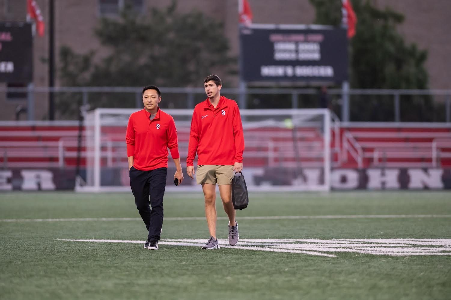
Dr. Jiang walking onto the field at a St. John's University men's soccer game
St. John's University men's soccer
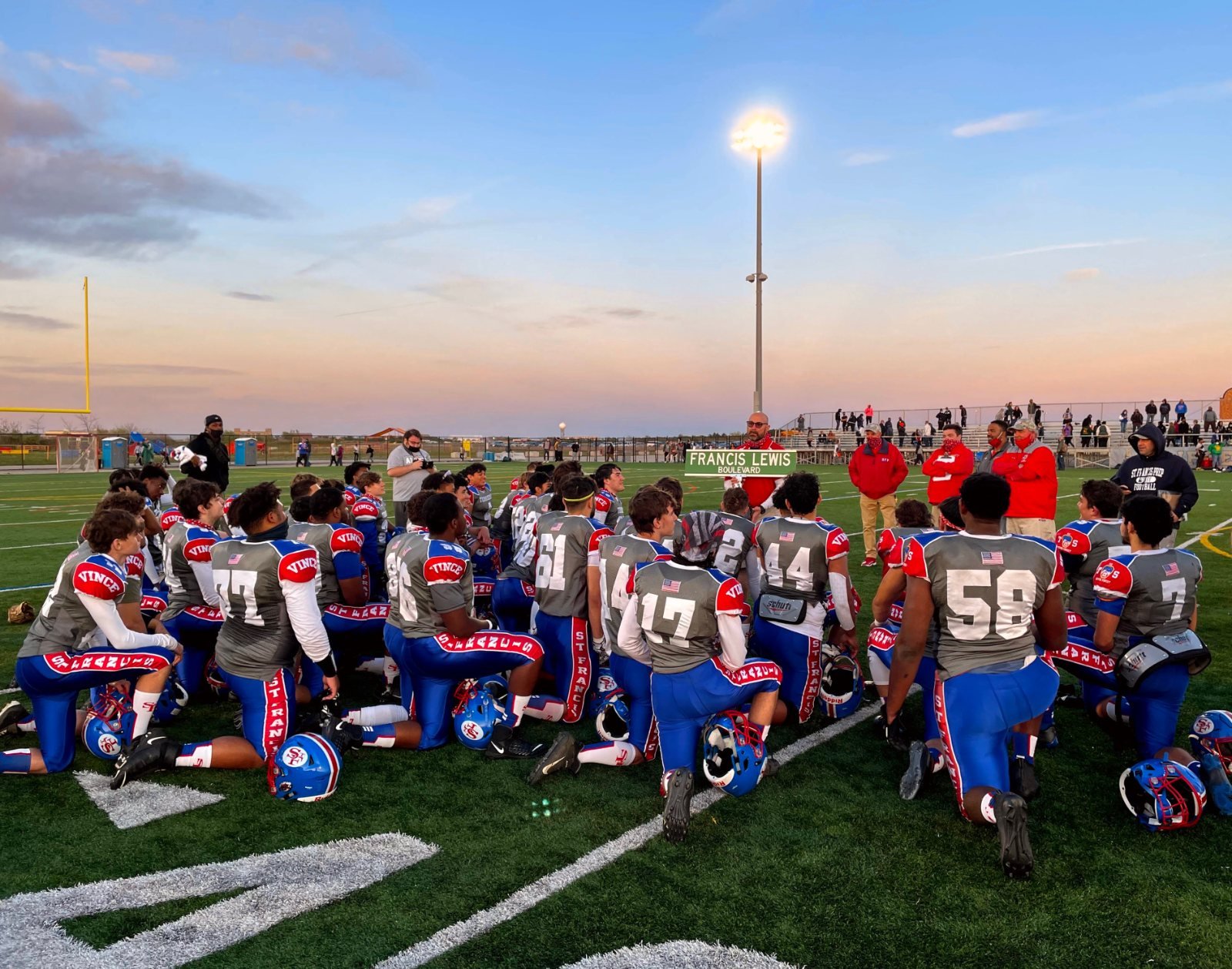
St. Francis Prep football team kneeling on the field after a game
St. Francis Prep football
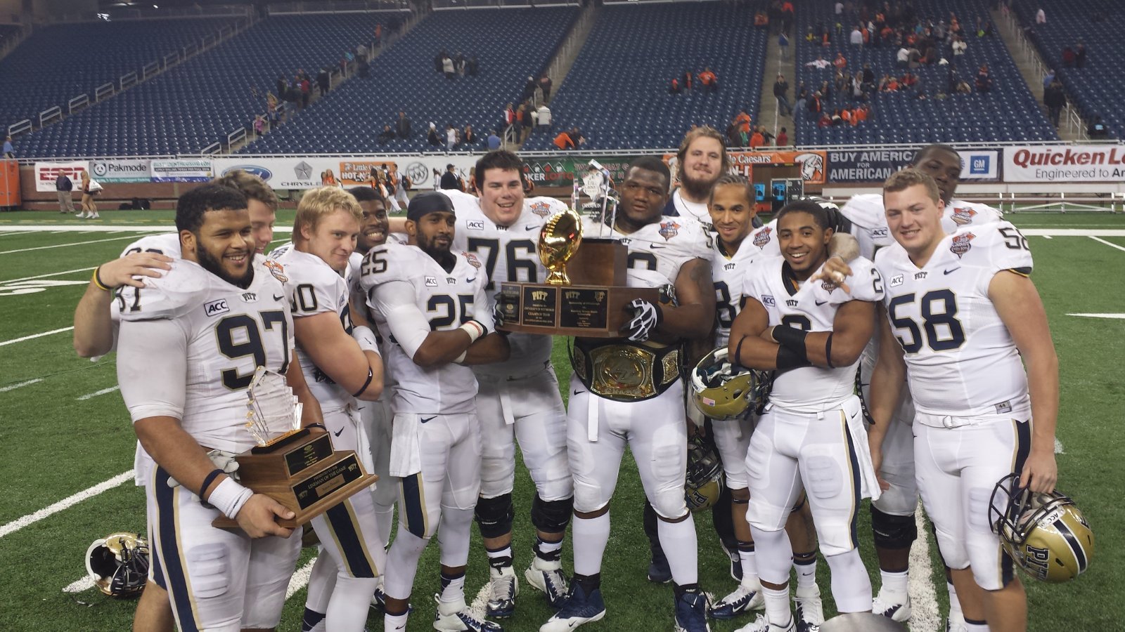
University of Pittsburgh football players celebrating with trophies on the field after the 2013 Little Caesars Pizza Bowl
University of Pittsburgh football, 2013 Little Caesars Pizza Bowl
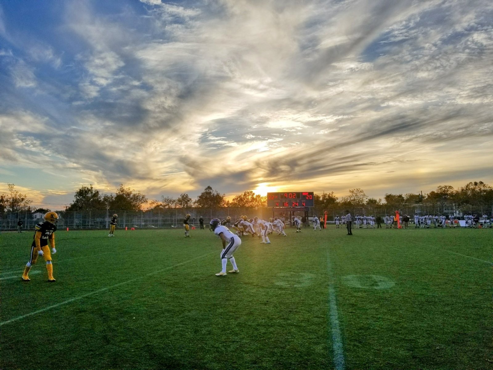
Holy Cross high school football game at sunset, viewed from the sideline
Holy Cross football
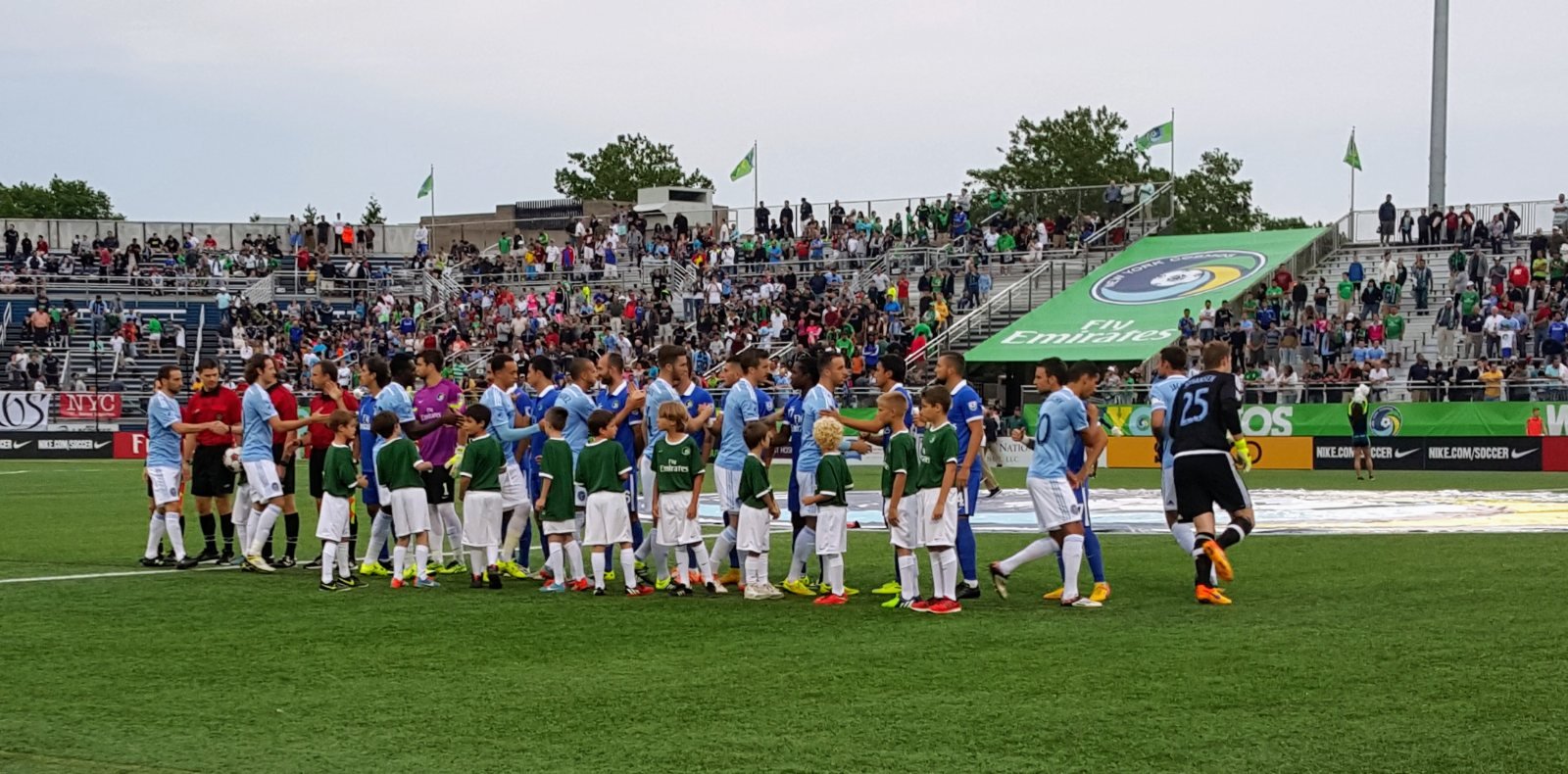
New York Cosmos and opposing players shaking hands with youth escorts before a professional soccer match
New York Cosmos, North American Soccer League
Athletes & active adultsAtletas y adultos activos运动员与活跃人群

Experienced with taking care of high-level athletes.Con experiencia en el cuidado de atletas de alto nivel.拥有照护高水平运动员的丰富经验。

Dr. Jiang treats pediatric, adolescent, and adult sports injuries. He has treated and operated on athletes at every level — from recreational to Division I to professional. Caring for an athlete involves more than the injury itself: the right decision weighs the athlete's goals, where they are in the season, and when it is truly safe to return to play. Dr. Jiang is currently a team physician for Holy Cross, St. Francis Prep, and Msgr. McClancy. He is also a U.S. Soccer Preferred Provider. From 2016 to 2024 he served as a team physician for St. John's University Athletics, covering Division I men's soccer, men's basketball, baseball, and lacrosse in the Big East. Dr. Jiang has also covered professional soccer with the New York Cosmos. As a sports medicine fellow, he was an assistant team physician for University of Pittsburgh football and basketball and provided care for the Pittsburgh Steelers.Atiende lesiones deportivas pediátricas y de adolescentes, además de las de atletas adultos, y ha operado a atletas de todos los niveles: desde recreativos hasta de División I y profesionales. Atender a un atleta implica más que la lesión en sí: la decisión correcta tiene en cuenta sus objetivos, el momento de la temporada y cuándo es realmente seguro volver a jugar. Ese criterio se adquiere con la experiencia. El Dr. Jiang es actualmente médico del equipo de Holy Cross, St. Francis Prep y Msgr. McClancy. También es proveedor médico preferido de U.S. Soccer. De 2016 a 2024 fue médico del equipo de St. John's University Athletics, cubriendo fútbol masculino, baloncesto masculino, béisbol y lacrosse de la División I en la Big East. También ha cubierto fútbol profesional con los New York Cosmos. Durante su subespecialidad (fellowship) en medicina deportiva, fue médico asistente del equipo de fútbol americano y baloncesto de la Universidad de Pittsburgh y brindó atención a los Pittsburgh Steelers.姜医生诊治儿童与青少年运动损伤,也照护成年运动员,手术对象涵盖各个水平的运动员——从业余爱好者到 NCAA 一级联赛乃至职业运动员。照护运动员不只是治疗损伤本身:正确的决定需要综合考虑运动员的目标、所处的赛季阶段,以及何时重返赛场才真正安全。这种判断力来自经验。姜医生目前担任 Holy Cross、St. Francis Prep 与 Msgr. McClancy 的队医。他也是美国足协(U.S. Soccer)认证的推荐医疗服务提供者。2016 至 2024 年,他担任 St. John's University Athletics 队医,负责大东联盟(Big East)NCAA 一级男子足球、男子篮球、棒球与长曲棍球。姜医生还曾为职业足球队 New York Cosmos 提供医疗服务。在运动医学专科培训期间,他担任匹兹堡大学橄榄球队与篮球队的助理队医,并为匹兹堡钢人队提供医疗服务。

{{ t.years }}
{{ t.org }}{{ t.org }}
{{ t.detail }}
Request an injury assessmentSolicitar una evaluación de lesión申请损伤评估
ExperienceExperiencia临床经验

We treat these injuries often.Tratamos estas lesiones con frecuencia.这些损伤,我们经常治疗。

Surgeons who treat an injury often tend to get better results. Dr. Jiang treats shoulder dislocation, ACL tears, and joint pain at a Los cirujanos que tratan una lesión con frecuencia suelen obtener mejores resultados. El Dr. Jiang trata la luxación de hombro, las roturas del LCA y el dolor articular con una 经常治疗某种损伤的医生,治疗效果往往更好。根据 Healthgrades 的治疗量数据,姜医生治疗肩关节脱位、前交叉韧带(ACL)撕裂与关节疼痛的very high frequencyfrecuencia muy alta频次远高于 relative to comparable surgeons, per Healthgrades treatment-volume data. en comparación con cirujanos similares, según los datos de volumen de tratamiento de Healthgrades.同类医生。

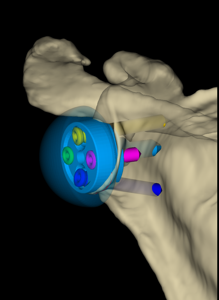
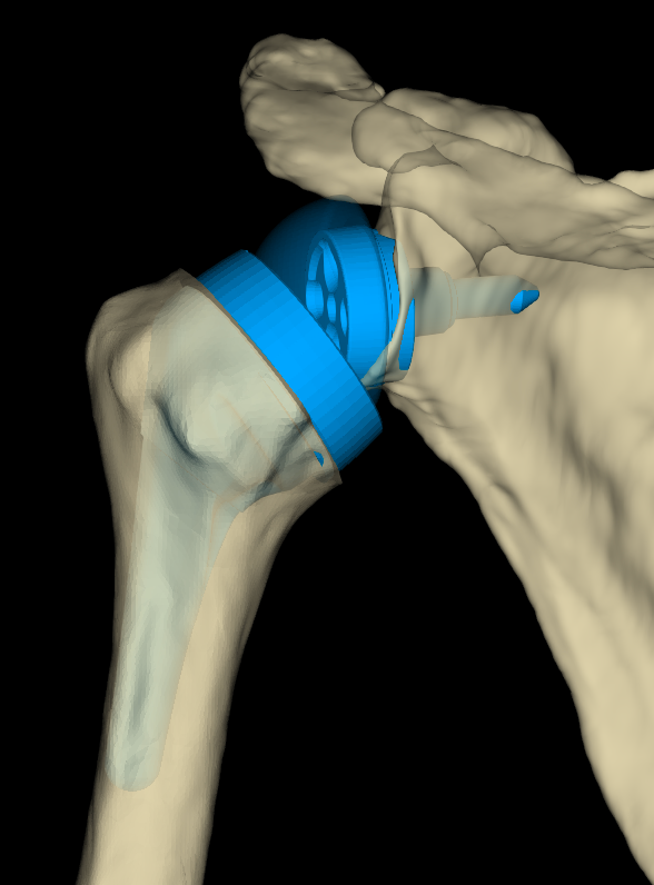
Core procedures: ACL reconstruction · arthroscopic rotator cuff repair · shoulder stabilization and labral repair · meniscus repair · multi-ligament knee reconstruction · total and reverse total shoulder replacement · cartilage repair · fracture fixation · PRP and orthobiologics.Procedimientos principales: reconstrucción del LCA · reparación artroscópica del manguito rotador · estabilización de hombro y reparación del labrum · reparación de menisco · reconstrucción multiligamentaria de rodilla · prótesis total y total invertida de hombro · reparación de cartílago · fijación de fracturas · PRP y ortobiológicos.核心手术:ACL 重建 · 关节镜下肩袖修补 · 肩关节稳定与盂唇修补 · 半月板修补 · 膝关节多韧带重建 · 全肩与反式全肩关节置换 · 软骨修复 · 骨折内固定 · PRP 与骨科生物制剂治疗。

{{ v.stat }}
{{ v.label }}

Multiple locations in QueensVarios consultorios en Queens皇后区多个诊疗地点

Imaging in the officeEstudios de imagen en el consultorio诊所内影像检查

X-ray, ultrasound, CT, and MRI are available at our offices. If necessary, depending on insurance, advanced imaging can be done the same day as your visit.Contamos con radiografías, ecografía, tomografía y resonancia magnética en nuestros consultorios. Si es necesario, y según su seguro, los estudios de imagen avanzados pueden hacerse el mismo día de su visita.我们的诊所配备 X 光、超声、CT 和核磁共振检查。如有需要,视您的保险而定,可在就诊当天完成高级影像检查。

{{ o.kicker }}
{{ o.name }}
{{ o.line1 }}
{{ o.line2 }}
{{ o.phone }}
{{ o.transit }}
Parking:Estacionamiento:停车: {{ o.parking }}
{{ o.hours }}

Patient experienceExperiencia de los pacientes患者评价

Aggregate ratings from independent platforms. Click each rating to read individual patient reviews.Valoraciones globales de plataformas independientes. Haga clic en cada valoración para leer las opiniones de los pacientes.来自独立平台的综合评分。点击各评分即可阅读患者的具体评价。

{{ p }}

Written testimonials will be published only with signed patient consent. Ratings above are summaries of third-party platform data and link to the original profiles. Ratings as of September 2026.Los testimonios escritos se publicarán únicamente con el consentimiento firmado del paciente. Las valoraciones anteriores resumen datos de plataformas externas y enlazan a los perfiles originales. Valoraciones actualizadas a septiembre de 2026.书面患者评价仅在获得患者签署同意后发布。以上评分为第三方平台数据的汇总,并链接至原始页面。评分截至 2026 年 9 月。

What brings you here?¿Qué le trae por aquí?您哪里不舒服?

Pick the area to read about the injury in plain language.Elija la zona para leer sobre la lesión en lenguaje sencillo.选择部位,用通俗的语言了解该损伤。
{{ area.kicker }}
{{ area.title }}
  • {{ it.name }}
Read about these →Leer más →了解详情 →

Get a clear answer about your injury, close to home.Reciba una respuesta clara sobre su lesión, cerca de casa.在家门口,得到关于伤情的明确答复。

Accepting new patients · Se habla español · 中文服务Aceptamos pacientes nuevos · Se habla español · 中文服务接受新患者 · Se habla español · 中文服务

Request an AppointmentSolicitar una cita预约就诊
(866) 670-6824
AboutSobre el Dr. Jiang关于姜医生

Kevin Jiang, MD

We help people recover from injury and get back to the things they care about, whether that's a sport, a job, or everyday life. First, we make sure you understand what is wrong. Then we decide on a treatment plan together. Often that means treatment without surgery, and surgery only when it is truly needed.Ayudamos a las personas a recuperarse de una lesión y volver a lo que les importa, ya sea un deporte, un trabajo o la vida diaria. Primero nos aseguramos de que usted entienda qué le pasa. Luego decidimos juntos el plan de tratamiento. Muchas veces no requiere cirugía, y recurrimos a ella solo cuando es realmente necesaria.我们帮助患者从损伤中康复,重新回到他们在意的事情上,无论是运动、工作还是日常生活。首先,我们会确保您理解问题所在;然后,我们一起决定治疗方案。很多情况下无需手术,只有在确实需要时才会考虑手术。

Dr. Jiang is a board-certified orthopaedic surgeon, fellowship-trained in sports medicine, specializing in minimally invasive and arthroscopic treatment of the shoulder, elbow, and knee. Dr. Jiang completed his orthopaedic surgery residency at Columbia University Medical Center, where he cared for Columbia, CUNY, and local high school athletes. Dr. Jiang then trained at the University of Pittsburgh's orthopaedic sports medicine program, working with Pitt football, the Pittsburgh Steelers, and the Pittsburgh Penguins.El Dr. Jiang es cirujano ortopédico certificado por la junta, con formación de subespecialidad en medicina deportiva, especializado en el tratamiento mínimamente invasivo y artroscópico del hombro, el codo y la rodilla. Completó su residencia en cirugía ortopédica en Columbia University Medical Center, donde atendió a atletas de Columbia, CUNY y escuelas secundarias locales. Después se formó en el programa de medicina deportiva ortopédica de la Universidad de Pittsburgh, trabajando con el fútbol americano de Pitt, los Pittsburgh Steelers y los Pittsburgh Penguins.姜医生是获得美国骨科委员会认证的骨科外科医生,接受过运动医学专科培训,专长于肩、肘、膝的微创与关节镜治疗。他在 Columbia University Medical Center 完成骨科住院医师培训,期间负责哥伦比亚大学、纽约市立大学以及当地高中运动员的诊疗。之后他在匹兹堡大学骨科运动医学项目接受培训,服务于匹兹堡大学橄榄球队、匹兹堡钢人队(Steelers)与匹兹堡企鹅队(Penguins)。

Dr. Jiang has published peer-reviewed articles and textbook chapters on shoulder instability, ACL injuries, knee ligament injuries, ankle fractures, and orthobiologics, and teaches as an Assistant Professor of Clinical Orthopaedic Surgery at both Weill Cornell Medical College and Columbia University Medical Center. Dr. Jiang spent eight years as a team physician for St. John's University Athletics and still covers high school athletics here in Queens.El Dr. Jiang ha publicado artículos revisados por pares y capítulos de libros sobre inestabilidad de hombro, lesiones del LCA, lesiones ligamentarias de rodilla, fracturas de tobillo y ortobiológicos, y enseña como Profesor Asistente de Cirugía Ortopédica Clínica tanto en Weill Cornell Medical College como en Columbia University Medical Center. Fue médico del equipo de St. John's University Athletics durante ocho años y sigue cubriendo deportes de secundaria aquí en Queens.姜医生在肩关节不稳、ACL 损伤、膝关节韧带损伤、踝关节骨折及骨科生物制剂等方向发表过多篇同行评审论文与专著章节,并在 Weill Cornell Medical College 与 Columbia University Medical Center 同时担任临床骨科外科助理教授。他曾担任 St. John's University Athletics 队医八年,如今仍在皇后区为高中体育赛事提供医疗保障。

Dr. Jiang sees patients in Jackson Heights and Long Island City, and operates and holds hospital clinic at NewYork-Presbyterian Queens in Flushing, where he is an attending in the Department of Orthopaedics and Rehabilitation. Dr. Jiang speaks Mandarin and Spanish.El Dr. Jiang atiende a pacientes en Jackson Heights y Long Island City, y opera y atiende en la clínica del hospital NewYork-Presbyterian Queens, en Flushing, donde es médico adjunto del Departamento de Ortopedia y Rehabilitación. Habla mandarín y español.姜医生在 Jackson Heights 与 Long Island City 出诊,在 Flushing 的 NewYork-Presbyterian Queens 进行手术并在医院门诊出诊,担任骨科与康复科主治医师。他会说普通话和西班牙语。
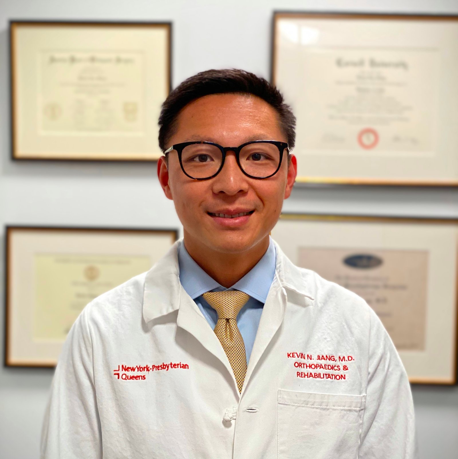

Dr. Kevin Jiang, MD, in a NewYork-Presbyterian Queens white coat, standing in front of his framed diplomas.
At a glanceEn resumen简要概览
Attending orthopaedic surgeon since 2014Cirujano ortopédico de planta desde 2014自 2014 年起担任骨科主治医师
Board-certified, American Board of Orthopaedic Surgery — with subspecialty certification in Orthopaedic Sports MedicineCertificado por el American Board of Orthopaedic Surgery, con certificación de subespecialidad en Medicina Deportiva Ortopédica美国骨科委员会认证,并具有骨科运动医学专科认证
Team Physician, high school athletics · St. John's University Athletics, 2016–2024Médico de equipo, deportes de secundaria · St. John's University Athletics, 2016–2024高中体育队医 · St. John's University Athletics 队医,2016–2024
English · Mandarin · SpanishInglés · Mandarín · Español英语 · 普通话 · 西班牙语
Accepting new patientsAceptando pacientes nuevos接受新患者
Hospital affiliationAfiliación hospitalaria医院隶属
NewYork-Presbyterian Queens Weill Cornell Medicine Columbia University Department of Orthopedic Surgery Hospital for Special Surgery
Professional societiesSociedades profesionales专业学会
American Board of Orthopaedic Surgery American Academy of Orthopaedic Surgeons American Orthopaedic Society for Sports Medicine
Institutional names denote affiliation and society names denote membership. Neither implies endorsement.Los nombres de las instituciones indican afiliación y los de las sociedades indican membresía. Ninguno implica respaldo.机构名称表示隶属关系,学会名称表示会员身份,均不代表其背书。
Athletes & active adultsAtletas y adultos activos运动员与活跃人群

Experienced with taking care of high-level athletes.Con experiencia en el cuidado de atletas de alto nivel.拥有照护高水平运动员的丰富经验。

Dr. Jiang treats pediatric, adolescent, and adult sports injuries. He has treated and operated on athletes at every level — from recreational to Division I to professional. Caring for an athlete involves more than the injury itself: the right decision weighs the athlete's goals, where they are in the season, and when it is truly safe to return to play. Dr. Jiang is currently a team physician for Holy Cross, St. Francis Prep, and Msgr. McClancy. He is also a U.S. Soccer Preferred Provider. From 2016 to 2024 he served as a team physician for St. John's University Athletics, covering Division I men's soccer, men's basketball, baseball, and lacrosse in the Big East. Dr. Jiang has also covered professional soccer with the New York Cosmos. As a sports medicine fellow, he was an assistant team physician for University of Pittsburgh football and basketball and provided care for the Pittsburgh Steelers.Atiende lesiones deportivas pediátricas y de adolescentes, además de las de atletas adultos, y ha operado a atletas de todos los niveles: desde recreativos hasta de División I y profesionales. Atender a un atleta implica más que la lesión en sí: la decisión correcta tiene en cuenta sus objetivos, el momento de la temporada y cuándo es realmente seguro volver a jugar. Ese criterio se adquiere con la experiencia. El Dr. Jiang es actualmente médico del equipo de Holy Cross, St. Francis Prep y Msgr. McClancy. También es proveedor médico preferido de U.S. Soccer. De 2016 a 2024 fue médico del equipo de St. John's University Athletics, cubriendo fútbol masculino, baloncesto masculino, béisbol y lacrosse de la División I en la Big East. También ha cubierto fútbol profesional con los New York Cosmos. Durante su subespecialidad (fellowship) en medicina deportiva, fue médico asistente del equipo de fútbol americano y baloncesto de la Universidad de Pittsburgh y brindó atención a los Pittsburgh Steelers.姜医生诊治儿童与青少年运动损伤,也照护成年运动员,手术对象涵盖各个水平的运动员——从业余爱好者到 NCAA 一级联赛乃至职业运动员。照护运动员不只是治疗损伤本身:正确的决定需要综合考虑运动员的目标、所处的赛季阶段,以及何时重返赛场才真正安全。这种判断力来自经验。姜医生目前担任 Holy Cross、St. Francis Prep 与 Msgr. McClancy 的队医。他也是美国足协(U.S. Soccer)认证的推荐医疗服务提供者。2016 至 2024 年,他担任 St. John's University Athletics 队医,负责大东联盟(Big East)NCAA 一级男子足球、男子篮球、棒球与长曲棍球。姜医生还曾为职业足球队 New York Cosmos 提供医疗服务。在运动医学专科培训期间,他担任匹兹堡大学橄榄球队与篮球队的助理队医,并为匹兹堡钢人队提供医疗服务。

Team & event coverageEquipos y eventos cubiertos队伍与赛事医疗保障
{{ t.years }}
{{ t.org }}{{ t.org }}
{{ t.detail }}
Dr. Jiang walking onto the field at a St. John's University men's soccer game
St. John's University men's soccer
St. Francis Prep football team kneeling on the field after a game
St. Francis Prep football
University of Pittsburgh football players celebrating with trophies on the field after the 2013 Little Caesars Pizza Bowl
University of Pittsburgh football, 2013 Little Caesars Pizza Bowl
Holy Cross high school football game at sunset, viewed from the sideline
Holy Cross football
New York Cosmos and opposing players shaking hands with youth escorts before a professional soccer match
New York Cosmos, North American Soccer League
Event coverageEventos cubiertos赛事医疗保障
  • {{ e }}

Philosophy of careFilosofía de atención诊疗理念

We help you understand the problem and the best options, and make a treatment plan together.Le ayudamos a entender el problema y las mejores opciones, y elaboramos juntos un plan de tratamiento.我们帮助您了解问题所在和最佳选择,并一起制定治疗方案。

{{ p.n }}

{{ p.text }}

CredentialsCredenciales资历

{{ c.label }}
{{ c.value }}

Research & teachingInvestigación y docencia科研与教学

Peer-reviewed articles and textbook chapters on shoulder instability, ACL injuries, knee ligament injuries, ankle fractures, and orthobiologics. Former clinical researcher at Columbia's Center for Orthopaedic Research and Center for Shoulder, Elbow, and Sports Medicine.Artículos revisados por pares y capítulos de libros sobre inestabilidad de hombro, lesiones del LCA, lesiones ligamentarias de rodilla, fracturas de tobillo y ortobiológicos. Fue investigador clínico en el Center for Orthopaedic Research y el Center for Shoulder, Elbow, and Sports Medicine de Columbia.在肩关节不稳、ACL 损伤、膝关节韧带损伤、踝关节骨折及骨科生物制剂方向发表同行评审论文与专著章节。曾任哥伦比亚大学骨科研究中心及肩肘与运动医学中心临床研究员。

Currently a sub-investigator on a Phase 3 multicenter trial evaluating an extended-release corticosteroid injection for glenohumeral osteoarthritis. Dr. Jiang helps train Hospital for Special Surgery (HSS) orthopaedic residents in trauma and sports medicine surgery, and the practice also teaches physician assistant students from St. John's University. Dr. Jiang was named NewYork-Presbyterian Queens Teacher of the Year in 2022.Actualmente es subinvestigador en un ensayo multicéntrico de Fase 3 que evalúa una inyección de corticosteroide de liberación prolongada para la artrosis glenohumeral. Participa en la formación de los residentes de ortopedia del Hospital for Special Surgery (HSS) en cirugía de trauma y de medicina deportiva, y el consultorio también recibe a estudiantes de asistente médico de St. John's University. En 2022 fue nombrado Profesor del Año de NewYork-Presbyterian Queens.目前担任一项三期多中心临床试验的分中心研究者,评估缓释型皮质类固醇注射治疗盂肱关节骨关节炎的效果。姜医生参与培训特种外科医院(HSS)的骨科住院医师,指导创伤与运动医学手术;诊所也接收 St. John's University 的医师助理专业学生。2022 年获评 NewYork-Presbyterian Queens 年度教师。

Selected publicationsPublicaciones seleccionadas代表性论著

Peer-reviewed articles and textbook chapters.Artículos revisados por pares y capítulos de libros.同行评审论文与专著章节。

{{ pub.cite }}
{{ pub.journal }}

Beyond the ORFuera del quirófano手术室之外

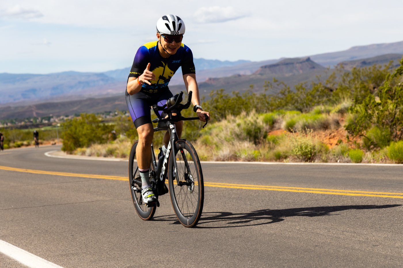
Dr. Jiang riding the bike leg of the St. George half Ironman, desert mountains behind him
Half Ironman, St. George, Utah

Dr. Jiang has finished a full Ironman and ten half-Ironmans. Dr. Jiang runs marathons, mountain bikes, road bikes, golfs, snowboards, and surfs.El Dr. Jiang ha completado un Ironman completo y diez medio Ironman. Corre maratones, practica ciclismo de montaña, ciclismo de ruta, golf, snowboard y surf.姜医生完成过一次全程铁人三项和十次半程铁人三项。他还跑马拉松,喜欢山地自行车、公路自行车、高尔夫、单板滑雪和冲浪。

Dr. Jiang grew up playing pickup basketball around New York City and went to Stuyvesant High School.Creció jugando partidos informales de baloncesto en canchas de toda la ciudad de Nueva York y estudió en Stuyvesant High School.他从小在纽约市各处的球场打街头篮球,毕业于 Stuyvesant 高中。

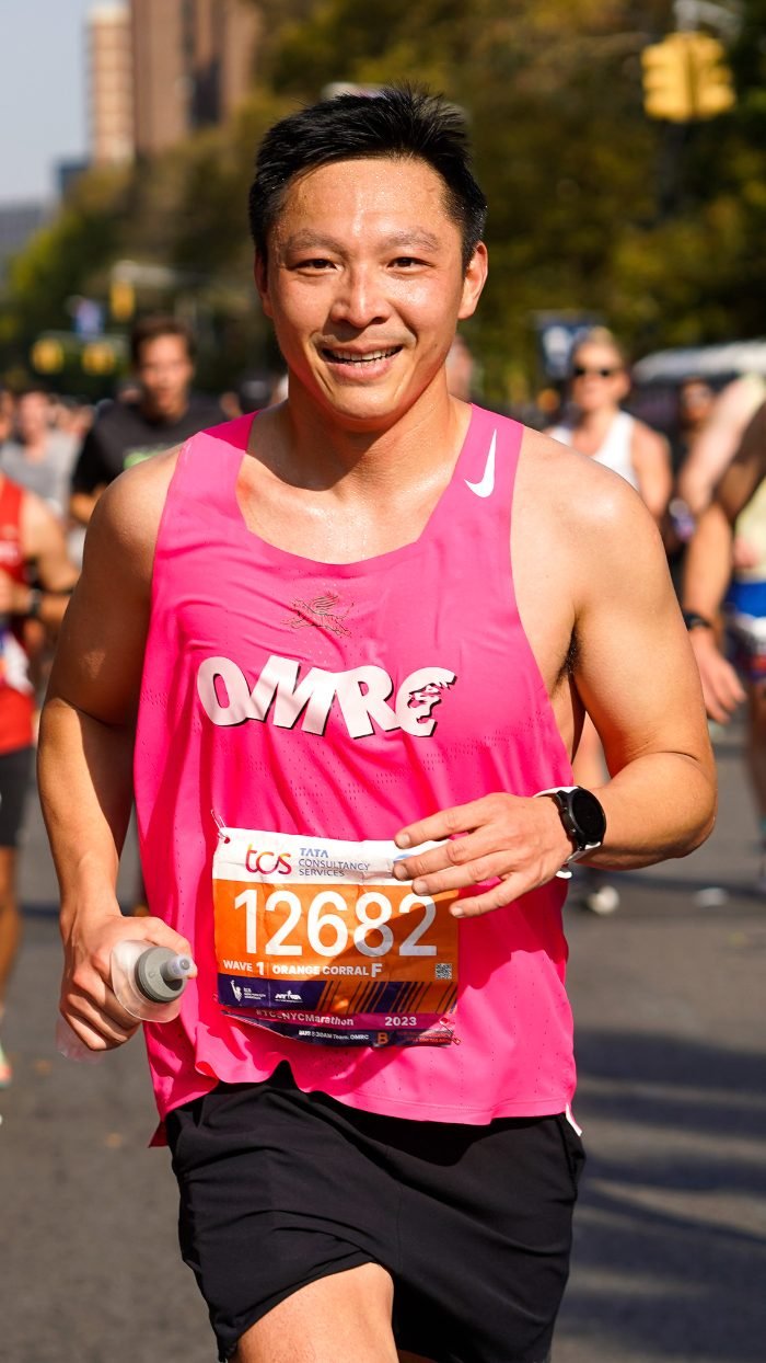
Dr. Jiang running the 2023 TCS New York City Marathon
NYC Marathon
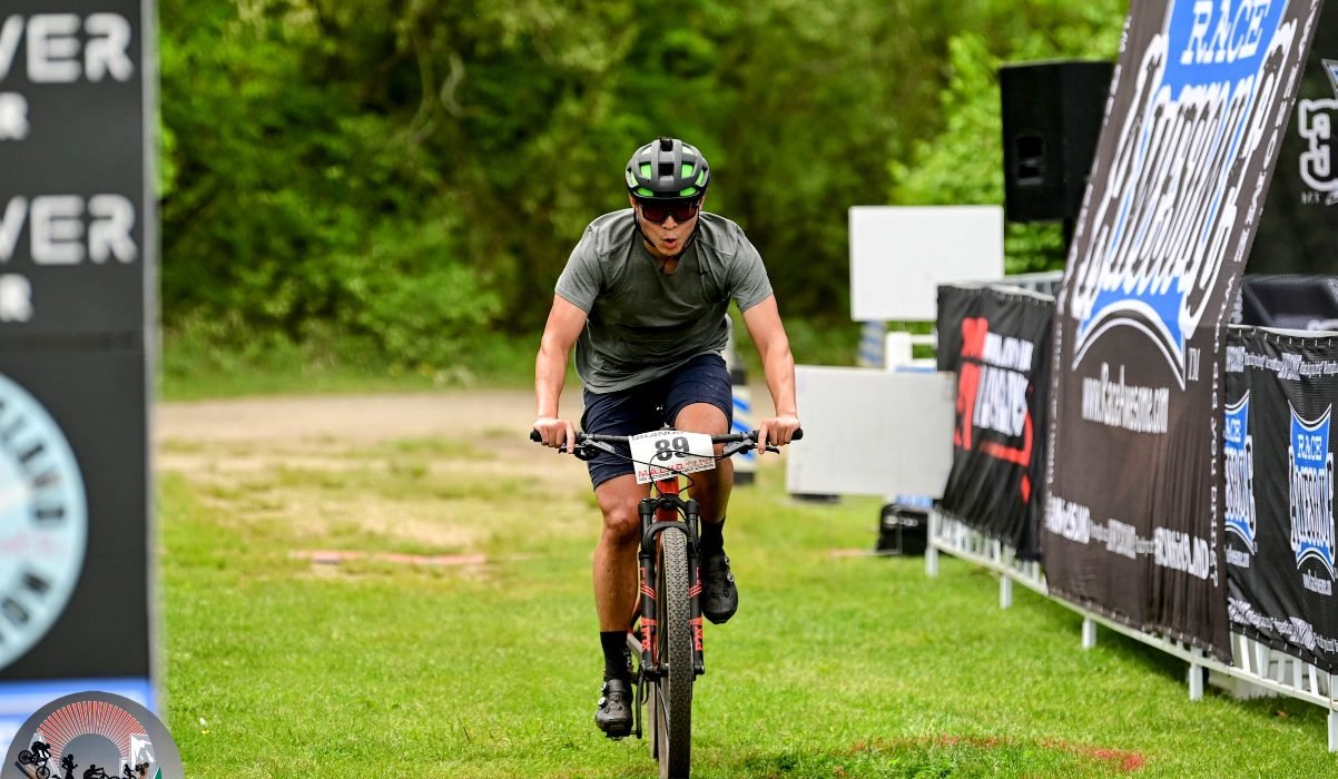
Dr. Jiang finishing a mountain bike race
Mountain biking
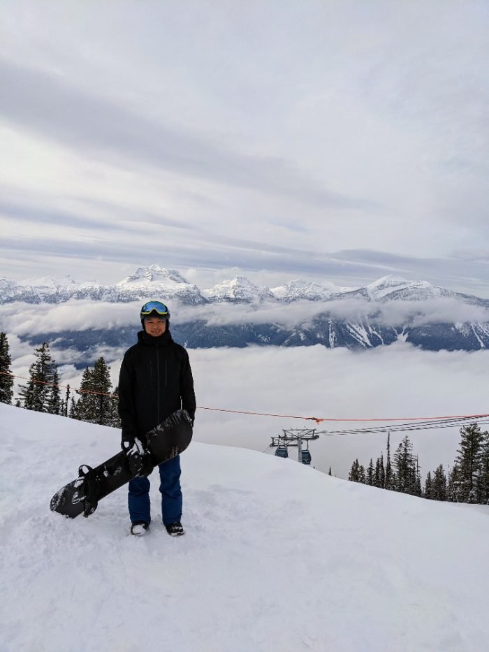
Dr. Jiang holding his snowboard on a mountain above the clouds
Snowboarding
In the newsEn las noticias媒体报道
Running the New York City Marathon with a patientCorrer el Maratón de Nueva York con un paciente与患者一起跑完纽约马拉松

Eight months after Dr. Jiang repaired his ruptured Achilles tendon, longtime distance runner Michael Bozzo ran the 2017 TCS New York City Marathon alongside him. They crossed the finish line together, injury free. Covered by ABC and the New York Daily News.Ocho meses después de que el Dr. Jiang le reparara el tendón de Aquiles roto, el corredor de fondo Michael Bozzo corrió junto a él el Maratón de Nueva York TCS 2017. Cruzaron la meta juntos, sin lesiones. Cubierto por ABC y el New York Daily News.姜医生为长跑爱好者 Michael Bozzo 修复断裂的跟腱八个月后,两人一起参加了 2017 年 TCS 纽约马拉松,并肩冲过终点,全程无伤。ABC 与《纽约每日新闻》对此进行了报道。

Read the story at NewYork-Presbyterian →Leer la historia en NewYork-Presbyterian →在 NewYork-Presbyterian 阅读全文 →

The teamEl equipo诊疗团队

You'll work with the same small team from your first visit through recovery.Trabajará con el mismo equipo pequeño desde su primera visita hasta su recuperación.从首次就诊到康复,您将始终与同一个小团队合作。

[ headshot ]
Alan Trang
Physician AssistantAsistente Médico医师助理

Works alongside Dr. Jiang in caring for patients — in clinic, around surgery, and through recovery.Trabaja junto al Dr. Jiang en el cuidado de los pacientes: en la consulta, alrededor de la cirugía y durante la recuperación.与姜医生一起照护患者——包括门诊、手术前后以及康复期间。

[ headshot ]
Sheleeza (Shelly) Hack
Practice CoordinatorCoordinadora de la consulta诊所协调员

Coordinates visits, books surgeries, prepares letters and forms, and handles the administrative side of your care.Coordina las visitas, programa las cirugías, prepara cartas y formularios, y se encarga de la parte administrativa de su atención.负责协调就诊、安排手术、处理信函与表格,并处理您诊疗中的各项行政事务。

Book a consult in Jackson Heights or Long Island City.Reserve una consulta en Jackson Heights o Long Island City.在 Jackson Heights 或 Long Island City 预约门诊。

Request an AppointmentSolicitar una cita预约就诊

Patient experienceExperiencia de los pacientes患者评价

Aggregate ratings from independent platforms. Click each rating to read individual patient reviews.Valoraciones globales de plataformas independientes. Haga clic en cada valoración para leer las opiniones de los pacientes.来自独立平台的综合评分。点击各评分即可阅读患者的具体评价。

{{ p }}

Written testimonials will be published only with signed patient consent. Ratings above are summaries of third-party platform data and link to the original profiles. Ratings as of September 2026.Los testimonios escritos se publicarán únicamente con el consentimiento firmado del paciente. Las valoraciones anteriores resumen datos de plataformas externas y enlazan a los perfiles originales. Valoraciones actualizadas a septiembre de 2026.书面患者评价仅在获得患者签署同意后发布。以上评分为第三方平台数据的汇总,并链接至原始页面。评分截至 2026 年 9 月。

{{ t.cond_en_note }}
← Back to ACL tear
ACL reconstruction · Evidence review

Choosing an ACL graft

When the ACL is reconstructed, it is replaced with a graft, a piece of tendon that becomes the new ligament. The graft can come from your own knee (an autograft) or from a donor (an allograft). Each option has strengths and trade-offs, and the right choice depends on your age, your sport, your work, and your goals. This page summarizes what the research shows, so you can make that decision with us.

The main options

Autograft
Quadriceps tendon
Taken from the tendon above the kneecap, with or without a small piece of bone.
Strengths
A large, thick graft with a predictable size. Recent randomized trials show failure rates similar to patellar tendon grafts, with less pain at the harvest site than the patellar tendon.5
Trade-offs
Some studies show slower recovery of quadriceps strength early on.
A strong option for most young, active patients, including those who kneel for work or sport.
Autograft
Patellar tendon (BTB)
The middle third of the tendon below the kneecap, with a small block of bone at each end.
Strengths
Bone-to-bone healing in the tunnels, and a long track record in high-level pivoting and contact athletes.
Trade-offs
More kneeling discomfort and front-of-knee pain than other grafts.7 Small risk of kneecap fracture.
Long the standard for high-level pivoting and contact athletes.
Donor tissue
Allograft
Tendon from a screened, sterilized tissue donor.
Strengths
No harvest from your own knee, so less early pain and a shorter operation.
Trade-offs
About four times the odds of graft rupture compared with autograft in young, active patients under 35,3 and the risk is highest in teenagers.4
In patients over 35, results are similar to autograft.13,14 Also used in some revision and multi-ligament cases.
Hamstring grafts, with a lateral extra-articular tenodesis
Hamstring tendon grafts were once widely used. We use them much less often now because the size of the harvested graft varies from person to person and can't be reliably predicted before surgery, and large registries show higher revision rates than with patellar tendon grafts.1,2 Hamstring grafts can still be used, but when we use one, we add a lateral extra-articular tenodesis (LET). In young, high-risk patients with hamstring grafts, adding an LET lowered graft rupture from 11% to 4%.9
Head to head

Quadriceps tendon vs patellar tendon

These are the two grafts we use most for young, active patients. Their results for stability, re-tear, and return to sport are similar. The main difference is how the front of the knee feels afterward.

Quadriceps tendon
Patellar tendon (BTB)
Graft failure
No significant difference in meta-analyses, including one limited to athletes.5,11
No significant difference from quadriceps tendon.5,11
Return to sport
Similar. In cutting and pivoting athletes, 88% returned to sport at 2 years.12
Similar. 83% returned in the same study.12
Kneeling & front-of-knee pain
Much less. Odds of anterior knee pain about 84% lower than with patellar tendon.11
More common, especially with kneeling.7,11
Pain during sport & work
Lower risk of moderate-to-severe kneecap symptoms (odds about 86% lower).11
Higher risk of kneecap symptoms during sport and work.11
Stability
Similar knee laxity on testing.5,11
Other trade-offs
Quadriceps strength can be slower to return early in rehab.
Small risk of kneecap fracture or patellar tendon problems.
Graft failure

How often does the new ligament tear?

Graft failure means the reconstructed ligament tears again or the knee becomes unstable enough to need a second (revision) surgery. Most people never have a graft failure, but the risk is not the same for everyone.

2.2% vs 2.5%
Revision at 2 years, quadriceps tendon vs patellar tendon, in the Swedish registry of more than 44,000 patients.6
4×
Higher odds of graft rupture with allograft than autograft in the MOON cohort.3
2.3×
Increase in the odds of graft rupture for every 10 years younger the patient is.3

Quadriceps tendon. In the Swedish registry of more than 44,000 patients, 2-year revision rates were similar: 2.2% for quadriceps tendon and 2.5% for patellar tendon.6 A 2026 meta-analysis of randomized trials found no difference in failure between quadriceps and patellar tendon grafts.5

Age and allograft. Younger age is the strongest single risk factor for re-tear. In patients 19 and younger, allograft carried nearly four times the risk of failure compared with autograft.4 For this reason, we generally avoid allograft in teenagers and young athletes.

Allograft in patients over 35. The picture is different in older, lower-demand patients. Reviews of the literature show that non-irradiated allografts give stability, function, and failure rates similar to autograft in skeletally mature, lower-risk patients,13,14 and patients over 40 who received an allograft had outcomes and revision rates similar to those who received their own tissue.14 For a recreational athlete in their mid-30s or older, an allograft can be a reasonable choice that avoids a harvest site and makes early recovery easier.

Kneeling & front-of-knee pain

Will it hurt to kneel?

Every graft leaves a small area where it was taken from, called the donor site. How much that area bothers you depends mostly on which graft was used.

21.3%
Severe kneeling difficulty at 2 years after patellar tendon graft.7
84% lower
Odds of front-of-knee pain with quadriceps tendon compared with patellar tendon.11
86% lower
Odds of moderate-to-severe kneecap symptoms during sport and work with quadriceps tendon.11

In the New Zealand ACL Registry of nearly 11,000 patients, about one in five had severe kneeling difficulty two years after a patellar tendon graft.7 Quadriceps tendon grafts cause much less front-of-knee and kneeling pain, in both meta-analyses and randomized trials.5,11 If you kneel for work, such as construction, plumbing, flooring, or childcare, or for sport, such as wrestling or catching, tell us. It matters for the choice.

Return to sport

Will I get back to my sport?

81%
Return to some sport after ACL reconstruction.8
65%
Return to their pre-injury level of sport.8
55%
Return to competitive sport.8

These averages come from a meta-analysis of 69 studies and more than 7,500 people.8 Return rates are higher in elite and professional athletes: in a 2026 review, more than 85% returned to play and almost 90% of those returned to their pre-injury level, at an average of about 9½ months.10

Graft choice has much less effect on returning to sport than strength, confidence, and completing rehabilitation. Return to play is decided by testing strength, hopping, and movement, not just by time since surgery.

Lowering re-tear risk

Lateral extra-articular tenodesis (LET)

For young patients at high risk of re-tear, a lateral extra-articular tenodesis (LET) can be added to the ACL reconstruction. It reinforces the outside of the knee against the twisting that the ACL controls.

11% → 4%
Graft rupture at 2 years, ACL reconstruction alone vs with LET, in patients aged 14 to 25.9
Similar
Return-to-sport activity levels with or without LET.9

These results come from the STABILITY randomized trial of more than 600 young, high-risk patients, all of whom had hamstring grafts.9 We consider LET for younger athletes in pivoting sports, knees that are very loose, and revision surgery.

How we choose

Your graft, your decision

There is no single best graft for everyone. At your visit, we will go through the factors that matter most for you:

  • Age and growth. Younger patients have the highest re-tear risk and do best with their own tissue. Children who are still growing need a technique that protects the growth plates.
  • Sport and level. Cutting, pivoting, and contact sports put more demand on the graft.
  • Kneeling. Work or sports that involve kneeling may favor a quadriceps tendon graft.
  • Prior surgery. A previous ACL reconstruction or graft harvest changes which options are available.
  • Other injuries. Meniscus tears and other ligament injuries can change the plan.
Request an Appointment
Back to ACL tear

References

  1. 1Gifstad T, Foss OA, Engebretsen L, et al. Lower risk of revision with patellar tendon autografts compared with hamstring autografts: a registry study based on 45,998 primary ACL reconstructions in Scandinavia. Am J Sports Med. 2014;42(10):2319–2328. Link →
  2. 2Persson A, Fjeldsgaard K, Gjertsen JE, et al. Increased risk of revision with hamstring tendon grafts compared with patellar tendon grafts after ACL reconstruction: a study of 12,643 patients from the Norwegian Cruciate Ligament Registry. Am J Sports Med. 2014;42(2):285–291. Link →
  3. 3Kaeding CC, Aros B, Pedroza A, et al. Allograft versus autograft anterior cruciate ligament reconstruction: predictors of failure from a MOON prospective longitudinal cohort. Sports Health. 2011;3(1):73–81. Link →
  4. 4Failure rates of autograft and allograft ACL reconstruction in patients 19 years of age and younger: a systematic review and meta-analysis. JBJS Open Access. 2020. Link →
  5. 5White et al. Quadriceps, hamstring and patella tendon autografts for primary ACL reconstruction demonstrate similar clinical outcomes, including graft failure, joint laxity and complications: a systematic review with meta-analysis of randomised controlled trials. Knee Surg Sports Traumatol Arthrosc. 2026. Link →
  6. 6Rizvanovic D, Waldén M, Forssblad M, et al. Patient-reported outcomes and revision rates after ACL reconstruction with quadriceps versus hamstring and patellar tendon autografts: sex-stratified results from the Swedish Knee Ligament Registry. Am J Sports Med. 2026;54(3):558–568. Link →
  7. 7Comparison of knee pain and difficulty with kneeling between patellar tendon and hamstring tendon autografts after ACL reconstruction: a study from the New Zealand ACL Registry. Orthop J Sports Med. Link →
  8. 8Ardern CL, Taylor NF, Feller JA, Webster KE. Fifty-five per cent return to competitive sport following anterior cruciate ligament reconstruction surgery: an updated systematic review and meta-analysis. Br J Sports Med. 2014;48(21):1543–1552. Link →
  9. 9Getgood AMJ, Bryant DM, Litchfield R, et al. Lateral extra-articular tenodesis reduces failure of hamstring tendon autograft ACL reconstruction: 2-year outcomes from the STABILITY study randomized clinical trial. Am J Sports Med. 2020;48(2):285–297. Link →
  10. 10D'Ambrosi R, et al. The majority of elite and professional athletes return to the preinjury level of activity after ACL reconstruction: a systematic review and meta-analysis. Knee Surg Sports Traumatol Arthrosc. 2026. Link →
  11. 11Zhang XF, Liu P, Huang JW, He YH. Efficacy and safety of quadriceps tendon autograft versus bone–patellar tendon–bone and hamstring tendon autografts for ACL reconstruction: a systematic review and meta-analysis. J Orthop Traumatol. 2024. Link →
  12. 12ACL reconstruction with quadriceps soft tissue autograft versus bone–patellar tendon–bone autograft in cutting and pivoting athletes: outcomes at minimum 2-year follow-up. Orthop J Sports Med. Link →
  13. 13Dhillon J, Kraeutler MJ, Belk JW, et al. Autograft and nonirradiated allograft for ACL reconstruction demonstrate similar clinical outcomes and graft failure rates: an updated systematic review. Arthrosc Sports Med Rehabil. 2022. Link →
  14. 14Paul BR, Robaina J, Parmar R, Carter T, Shah A. Myths and facts about allograft use in anterior cruciate ligament reconstruction: a detailed review of the literature. JBJS Rev. 2025;13(4):e25.00013. Link →

This page summarizes published research for patient education. Results from studies describe groups of patients and may not apply to you. It is not a substitute for an evaluation and a discussion of your own knee.

{{ t.cond_en_note }}
← Back to meniscus tear
Meniscus tears · Evidence review

Meniscus tears in a knee with arthritis

If you are over 40 and have knee pain, an MRI will often show a meniscus tear. In a knee that also has arthritis, that tear is usually part of the wear in the joint, not a separate injury that needs to be fixed. This page explains what the research shows, why we start with treatment that doesn't involve surgery, and why arthroscopy is our last option in this setting rather than our first.

Tears are common

A tear on MRI is not always the source of pain

The meniscus wears with age, just like cartilage. Degenerative tears are very common in middle-aged and older adults, including people who have no knee pain at all.

61%
Of people with a meniscus tear on MRI had no knee pain, aching, or stiffness in the previous month.1
60%
Prevalence of meniscus tears in people with knee arthritis who had no knee symptoms.1
63%
Prevalence in people with knee arthritis who did have symptoms. Nearly the same.1

In this large community study, tears were almost as common in people without pain as in people with pain once arthritis was present.1 That is why we treat you and your symptoms, not the MRI.

What the trials show

Arthroscopy does not outperform non-surgical care

Several high-quality randomized trials have compared arthroscopic surgery with physical therapy, exercise, or a sham (placebo) operation in degenerative knees.

METEORNEJM 2013
330 patients with a meniscus tear and mild-to-moderate arthritis were assigned to partial meniscectomy plus physical therapy, or physical therapy alone. At 6 and 12 months, function improved similarly in both groups. About 30% of the therapy group chose surgery within 6 months, and they did well too, so trying therapy first did not cost them anything.2
FIDELITYNEJM 2013
In patients with a degenerative medial meniscus tear and no arthritis, partial meniscectomy was compared with a sham operation in which the surgeon only made the incisions. Both groups improved by the same amount at 12 months.3
Kise et al.BMJ 2016
A 12-week supervised exercise program was compared with partial meniscectomy in patients with degenerative tears. At 2 years, there was no clinically meaningful difference in outcome, and the exercise group had better thigh-muscle strength at 3 months.4
Kirkley et al.NEJM 2008
In patients with knee arthritis, arthroscopic washout and debridement added to physical and medical therapy gave no additional benefit over therapy alone at 2 years.5
Moseley et al.NEJM 2002
Arthroscopic washout or debridement for knee arthritis was compared with a sham operation. Outcomes were no better than placebo at any point over 2 years.6

Based on this body of evidence, an international panel that included orthopaedic surgeons issued a strong recommendation against arthroscopic surgery for nearly all patients with degenerative knee disease, with or without a meniscus tear, mechanical symptoms, or arthritis on X-ray.7

Why it can make things worse

Removing meniscus raises the load on the cartilage

The meniscus is a shock absorber that shares the load across the joint. Removing part of it concentrates force on the cartilage that is already worn.

~3×
Higher risk of eventual knee replacement after arthroscopic surgery for a degenerative meniscus tear.8

In some knees, a rare but serious problem called subchondral insufficiency fracture can develop after arthroscopy. It is a collapse of the bone under the cartilage, which causes a sudden increase in pain in the weeks after surgery and can speed up the need for knee replacement.9 There are also the general risks of any surgery, such as infection, blood clots, and stiffness, for a procedure that, on average, does not give better results.

What we do instead

Treatment that works for degenerative knees

  • Exercise and physical therapy. Strengthening the quadriceps, hips, and core is the single most effective treatment, and it is the foundation of every plan.2,4
  • Weight management. Every pound of body weight puts several pounds of force across the knee with each step, so even modest weight loss reduces pain.
  • Medication. Anti-inflammatory medicine, by mouth or as a topical gel, when it is safe for you.
  • Injections. Corticosteroid, hyaluronic acid, or orthobiologic (PRP) injections can calm a flare and make it easier to stay active.
  • Bracing. An unloader brace shifts weight away from the worn side of the knee.
  • Realignment. In younger, active patients with bowlegged or knock-kneed alignment, an osteotomy can move the load to the healthier side of the knee.
  • Knee replacement. When arthritis is advanced and other treatments no longer help, partial or total knee replacement is the treatment that reliably relieves pain.
When arthroscopy is considered

The last option, for specific problems

Arthroscopy still has a role, but in a knee with arthritis it is reserved for specific situations, and only after a full course of non-surgical treatment:

  • A truly locked knee. A large flap or bucket-handle fragment that is physically blocking the knee from straightening, rather than the catching or clicking that is common with arthritis.
  • A new injury on top of mild wear. A clear twisting injury with a fresh, unstable tear, where the arthritis is minimal.
  • Repairable tears. Some tears, such as root tears in knees with little arthritis, can be repaired rather than removed. That is a different operation with a different goal: saving the meniscus, not trimming it.

Even then, we will talk through the expected benefit, the risks, and the alternatives, so the decision is yours and is made with the evidence in front of you.

Request an Appointment
Back to meniscus tear

References

  1. 1Englund M, Guermazi A, Gale D, et al. Incidental meniscal findings on knee MRI in middle-aged and elderly persons. N Engl J Med. 2008;359(11):1108–1115. Link →
  2. 2Katz JN, Brophy RH, Chaisson CE, et al. Surgery versus physical therapy for a meniscal tear and osteoarthritis (METEOR). N Engl J Med. 2013;368(18):1675–1684. Link →
  3. 3Sihvonen R, Paavola M, Malmivaara A, et al. Arthroscopic partial meniscectomy versus sham surgery for a degenerative meniscal tear (FIDELITY). N Engl J Med. 2013;369(26):2515–2524. Link →
  4. 4Kise NJ, Risberg MA, Stensrud S, et al. Exercise therapy versus arthroscopic partial meniscectomy for degenerative meniscal tear in middle aged patients: randomised controlled trial with two year follow-up. BMJ. 2016;354:i3740. Link →
  5. 5Kirkley A, Birmingham TB, Litchfield RB, et al. A randomized trial of arthroscopic surgery for osteoarthritis of the knee. N Engl J Med. 2008;359(11):1097–1107. Link →
  6. 6Moseley JB, O’Malley K, Petersen NJ, et al. A controlled trial of arthroscopic surgery for osteoarthritis of the knee. N Engl J Med. 2002;347(2):81–88. Link →
  7. 7Siemieniuk RAC, Harris IA, Agoritsas T, et al. Arthroscopic surgery for degenerative knee arthritis and meniscal tears: a clinical practice guideline. BMJ. 2017;357:j1982. Link →
  8. 8Rongen JJ, Rovers MM, van Tienen TG, et al. Increased risk for knee replacement surgery after arthroscopic surgery for degenerative meniscal tears: a multi-center longitudinal observational study using data from the Osteoarthritis Initiative. Osteoarthritis Cartilage. 2017;25(1):23–29. Link →
  9. 9Hussain ZB, Chahla J, Mandelbaum BR, Gomoll AH, LaPrade RF. The role of meniscal tears in spontaneous osteonecrosis of the knee: a systematic review of suspected etiology and a call to revisit nomenclature. Am J Sports Med. 2019;47(2):501–507. Link →

This page summarizes published research for patient education. Study results describe groups of patients and may not apply to you. It is not a substitute for an evaluation and a discussion of your own knee.

{{ t.cond_en_note }}
← Back to rotator cuff tear
Rotator cuff repair · Evidence review

Rotator cuff repair: healing, recovery, and results

Rotator cuff repair reattaches the torn tendon to the bone, and the body then has to heal it there. How well that happens depends on the size of the tear, the quality of the tendon, your age and health, and how carefully the repair is protected while it heals. This page summarizes what the research shows, so you know what to expect.

Healing rates

How often does the tendon heal?

In research studies, healing is checked with ultrasound or MRI after surgery. In everyday care, imaging is only done if there is persistent pain or weakness. A "retear" means the tendon did not stay attached, either fully or in part. The biggest single factor is the size of the tear.

8.7%
Retear in small tears (1 cm or less) at 6 months.1
14.7%
Retear in medium tears (1 to 3 cm).1
44.8%
Retear in large tears (3 to 5 cm).1

These figures come from more than 1,100 arthroscopic repairs.1 Across more than 8,000 patients in a large meta-analysis, about one in four repairs did not fully heal.2 In chronic massive tears, the retear rate is much higher, but pain still dropped from an average of 5.9 to 1.7 out of 10, and motion and function improved substantially.3 This is one reason we recommend repairing a tear before it becomes large.

Age

Does age matter?

95%
Healing of isolated supraspinatus repairs in patients under 55.4
76%
Healing in patients aged 55 to 64.4
43%
Healing in patients 65 and older.4

Tendons heal less reliably with age.4 But healing on an MRI is not the same as a good result. In patients over 70, satisfaction after arthroscopic repair was about 95%,5 and in one comparison, patients over 70 gained more function than patients under 50, despite lower healing rates.6 Age alone is not a reason to avoid repair.

Risk factors

What raises the risk of a retear?

  • Tear size and retraction. Larger, more retracted tears are the strongest predictor of retear.1,7
  • Muscle quality. When the muscle has turned to fat (fatty infiltration), the risk of retear roughly triples.7
  • Smoking. Smokers have about twice the risk of retear and a higher risk of needing another operation.8
  • Diabetes and high cholesterol. Both are linked to higher retear risk.7
  • Traumatic tears do better. A tear from a clear injury, such as a fall or lifting something heavy, usually happens in a healthier tendon with healthier muscle than a tear that develops slowly from wear.9 The advantage depends on timing: one large study found that traumatic and wear-related tears healed at similar rates overall.15
  • Waiting too long. For tears from an injury, results are better when the repair is done within a few months of the injury.9

Several of these are things you can change. Stopping smoking before surgery, and good control of blood sugar and cholesterol, give the repair a better chance to heal.

Clinical results

If it retears, does it matter?

Most patients do well after rotator cuff repair, including many whose repair does not fully heal. In a meta-analysis of more than 3,000 repairs, every outcome, including pain, function, and quality of life, improved substantially from before surgery, whether or not the tendon healed.10

Better
Strength, motion, and function scores when the tendon heals.10,11
Similar
Quality of life with or without a retear.10

A healed repair does give better results than a retear, especially for strength.10,11 That is why we focus on giving the tendon the best chance to heal, with careful surgical technique and a rehabilitation plan that protects the repair.

Recovery

What recovery looks like

Tendon-to-bone healing takes months, and the repair is at its weakest in the first several weeks. Rehabilitation is designed to protect it while keeping the shoulder from getting stiff.

Weeks 0–6
Sling, with gentle passive motion guided by your therapist. No active lifting.
Weeks 6–12
Sling weaned. Active motion begins and light daily use returns.
Months 3–6
Progressive strengthening.
Months 6–12
Return to overhead sport and heavy work. Strength keeps improving for up to a year.

Randomized trials show that starting gentle motion early improves range of motion without increasing the retear rate,12,13 although some reviews suggest a more protected approach for larger tears.14 Your timeline is tailored to the size and quality of your repair.

What this means for you

Making the decision together

Smaller tears heal best, and tears tend to get larger over time. For an active patient with a painful tear, repairing it while it is still small to medium gives the best chance of healing. For larger tears, we will talk honestly about the healing rate, and about other options such as superior capsular reconstruction, tendon transfer, or reverse shoulder replacement when a tear can't be repaired.

Request an Appointment
Back to rotator cuff tear

References

  1. 1Does tear size influence factors associated with early retear, satisfaction, and functional outcomes after arthroscopic rotator cuff repair? PLOS One. 2026. Link →
  2. 2McElvany MD, McGoldrick E, Gee AO, Neradilek MB, Matsen FA 3rd. Rotator cuff repair: published evidence on factors associated with repair integrity and clinical outcome. Am J Sports Med. 2015;43(2):491–500. Link →
  3. 3Henry P, Wasserstein D, Park S, et al. Arthroscopic repair for chronic massive rotator cuff tears: a systematic review. Arthroscopy. 2015;31(12):2472–2480. Link →
  4. 4Boileau P, Brassart N, Watkinson DJ, et al. Arthroscopic repair of full-thickness tears of the supraspinatus: does the tendon really heal? J Bone Joint Surg Am. 2005;87(6):1229–1240. Link →
  5. 5Arthroscopic rotator cuff repair in patients over 70 years of age: a systematic review. J Orthop Traumatol. 2021. Link →
  6. 6Hawkins Award 2023: clinical outcome and tendon healing after arthroscopic repair of isolated supraspinatus tears in patients older than 70 years vs younger than 50 years. J Shoulder Elbow Surg. 2024. Link →
  7. 7Incidence and risk factors for rotator cuff retear after arthroscopic rotator cuff repair in middle-aged and older patients: a systematic review and meta-analysis. BMC Musculoskelet Disord. 2026. Link →
  8. 8The effects of smoking on clinical and structural outcomes after rotator cuff repair: a systematic review and meta-analysis. J Shoulder Elbow Surg. 2022. Link →
  9. 9Mall NA, Lee AS, Chahal J, et al. An evidenced-based examination of the epidemiology and outcomes of traumatic rotator cuff tears. Arthroscopy. 2013;29(2):366–376. Link →
  10. 10The clinical impact of retears after repair of posterosuperior rotator cuff tears: a systematic review and meta-analysis. J Shoulder Elbow Surg. 2023. Link →
  11. 11Yang J Jr, Robbins M, Reilly J, Maerz T, Anderson K. The clinical effect of a rotator cuff retear: a meta-analysis of arthroscopic single-row and double-row repairs. Am J Sports Med. 2017. Link →
  12. 12Chan K, MacDermid JC, Hoppe DJ, et al. Delayed versus early motion after arthroscopic rotator cuff repair: a meta-analysis. J Shoulder Elbow Surg. 2014. Link →
  13. 13Early versus delayed mobilization for arthroscopic rotator cuff repair (small to large sized tear): a meta-analysis of randomized controlled trials. BMC Musculoskelet Disord. 2023. Link →
  14. 14Houck DA, Kraeutler MJ, Schuette HB, McCarty EC, Bravman JT. Early versus delayed motion after rotator cuff repair: a systematic review of overlapping meta-analyses. Am J Sports Med. 2017. Link →
  15. 15Guevara-Alvarez A, et al. Traumatic and atraumatic rotator cuff tears have the same rates of healing. Arthrosc Sports Med Rehabil. 2024. Link →

This page summarizes published research for patient education. Study results describe groups of patients and may not apply to you. It is not a substitute for an evaluation and a discussion of your own shoulder.

{{ t.cond_en_note }}
← Back to distal biceps rupture
Distal biceps rupture · Evidence review

Distal biceps rupture: repair or not?

The biceps does two jobs at the elbow: it bends the elbow (flexion) and, more importantly, it turns the palm up (supination), the motion you use to turn a key, a screwdriver, or a doorknob. When the tendon tears off the forearm bone, the arm still works, but it is weaker. This page explains how much strength is lost without surgery, what repair restores, and the trade-offs of each approach.

What is lost

Strength without the biceps attached

Other muscles can still bend the elbow, so many people are surprised that they can use the arm soon after the injury. The biceps is the main muscle that turns the palm up, though, so twisting strength suffers most.

~40%
Loss of supination (palm-up twisting) strength without repair.1
~30%
Loss of elbow flexion strength without repair.1

These figures come from Morrey and colleagues' classic study, which also found that early reattachment restored normal flexion and supination strength, while late reattachment improved strength without fully restoring it.1 Supination endurance is also reduced, so the arm tires quickly with repeated twisting, lifting, or tool use.2

Surgery vs no surgery

What the studies show

Supination strength (% of other arm)
Repair
89%
No repair
63%
Flexion strength (% of other arm)
Repair
95%
No repair
83%

Pooled results from a systematic review of 53 studies and 1,380 patients.3

A 2022 meta-analysis of 62 studies and nearly 2,500 cases found that surgery gave about 26% more flexion strength, 28% more supination strength, and 34% more supination endurance than non-surgical treatment, along with better arm-function scores.2

Non-surgical treatment is still a reasonable option for some people. In one study, patients treated without surgery kept a median of 93% of their flexion strength and 63% of their supination strength, and many were satisfied with their result.4 It can be a good choice for people with lower physical demands, those who mainly use the other arm, or those with health problems that make surgery risky. Without repair, some people also notice cramping in the upper arm and a change in the shape of the biceps.

Timing

Why sooner is better

After the tendon tears, it pulls back up the arm and begins to scar in place. In the first few weeks, it can usually be brought back to the bone and repaired directly. After that, the tendon may be too short or scarred to reach, and a tendon graft may be needed to bridge the gap. Early repair restores strength more fully than late repair.1 If you think you have torn your biceps, it is worth being seen within a week or two.

Our approach

Two-incision repair with small buttons

Dr. Jiang uses a two-incision technique: a small incision at the front of the elbow to retrieve the tendon, and a second small incision on the back of the forearm to place it back on its natural attachment point on the radius bone. The tendon is held in place with small buttons.

In a randomized trial of 90 patients comparing single-incision and two-incision repairs, function and pain were similar. The two-incision group regained slightly more flexion strength (104% vs 94% of the other arm) and had fewer minor complications, mainly less temporary numbness of the forearm.5 In a review of 784 repairs, serious complications were uncommon with either approach.6

Possible complications
  • Temporary numbness on the outside of the forearm from irritation of a skin nerve. This is the most common issue and usually goes away on its own.5,7
  • Extra bone formation (heterotopic ossification) near the repair, which can limit forearm rotation. It is more common with two-incision repair and rarely needs treatment.6,7
  • Nerve weakness in the wrist or fingers is rare, and most cases recover.6
  • Re-rupture is uncommon and usually linked to a fall or heavy lifting too early.5
Recovery

What to expect after repair

  • First 2 weeks. A splint or brace to protect the repair.
  • Weeks 2–6. Gentle motion in a hinged brace. No lifting with the arm.
  • Weeks 6–12. Brace off, full motion, and light strengthening.
  • Months 3–6. Progressive strengthening and a return to heavy work, lifting, and sport.
Request an Appointment
Back to distal biceps rupture

References

  1. 1Morrey BF, Askew LJ, An KN, Dobyns JH. Rupture of the distal tendon of the biceps brachii: a biomechanical study. J Bone Joint Surg Am. 1985;67(3):418–421. Link →
  2. 2Operative vs nonoperative treatment of distal biceps ruptures: a systematic review and meta-analysis. J Shoulder Elbow Surg. 2022. Link →
  3. 3Cuzzolin M, Secco D, Guerra E, et al. Operative versus nonoperative management for distal biceps brachii tendon lesions: a systematic review and meta-analysis. Orthop J Sports Med. 2021;9(10). Link →
  4. 4Freeman CR, McCormick KR, Mahoney D, Baratz M, Lubahn JD. Nonoperative treatment of distal biceps tendon ruptures compared with a historical control group. J Bone Joint Surg Am. 2009;91(10):2329–2334. Link →
  5. 5Grewal R, Athwal GS, MacDermid JC, et al. Single versus double-incision technique for the repair of acute distal biceps tendon ruptures: a randomized clinical trial. J Bone Joint Surg Am. 2012;94(13):1166–1174. Link →
  6. 6Ford SE, Andersen JS, Macknet DM, et al. Major complications after distal biceps tendon repairs: an analysis of complications in 784 surgical repairs. J Shoulder Elbow Surg. 2018. Link →
  7. 7Amin NH, Volpi A, Lynch TS, et al. Complications of distal biceps tendon repair: a meta-analysis of single-incision versus double-incision surgical technique. Orthop J Sports Med. 2016. Link →

This page summarizes published research for patient education. Study results describe groups of patients and may not apply to you. It is not a substitute for an evaluation and a discussion of your own elbow.

{{ t.cond_en_note }}
← Back to Achilles tendon rupture
Achilles tendon rupture · Evidence review

Achilles rupture: surgery or no surgery?

A ruptured Achilles tendon can heal with surgery or without it. Large randomized trials now show that both approaches give similar overall function at one year. The differences are in the risk of tearing it again, the risks of surgery itself, and how much calf strength comes back. This page lays out those trade-offs so you can choose the right path with us.

The big picture

How the two approaches compare

Surgical repair
Non-surgical treatment
Function at 1 year
Similar.1
Similar.1
Re-rupture
Lower: 0.6% in the largest trial.1
Higher: 6.2% in the same trial.1
Calf strength
Smaller strength deficit in some studies.4
Larger strength deficit in some studies.4
Complications
More: infection, wound problems, and nerve irritation.1,2
Fewer. Blood clots are the most common.2
Return to work
Earlier, especially after minimally invasive repair.3
Later on average.3
Re-rupture

How often does it tear again?

0.6%
Re-rupture after surgical repair, open or minimally invasive, in a 554-patient randomized trial.1
6.2%
Re-rupture after non-surgical treatment in the same trial.1
2.3% vs 3.9%
Re-rupture with surgery vs without across 29 studies and more than 15,000 patients.2

Every major study agrees that surgery lowers the chance of re-rupture.1,2,3 How big that difference is depends a lot on rehabilitation. When non-surgical treatment uses an accelerated functional rehab program, with early weight-bearing in a boot and early ankle motion, the difference in re-rupture becomes small and is no longer statistically significant.2 In a randomized trial of 144 patients using this kind of rehab, there were 2 re-ruptures with surgery and 3 without.5 A re-rupture is a real setback: it usually means surgery anyway, with a longer recovery.

Strength

How much calf strength comes back?

The Achilles is the tendon that lets you push off, climb stairs, run, and jump. After a rupture, the tendon can heal slightly longer than before, which reduces push-off power and how high you can rise up on your toes.

Some calf weakness and a lower single-leg heel rise often remain with either treatment.1 Studies differ on how much surgery helps. In the large Norwegian trial and the accelerated-rehab trial, physical performance was similar between groups.1,5 Other studies found a smaller strength deficit after repair.1,4 For sprinting, jumping, and cutting sports, even a small difference in push-off power can matter.

Risks of surgery

Complications to know about

4.9% vs 1.6%
Overall complication rate with surgery vs without.2
2.8%
Infection rate after surgery, the most common surgical complication.2
2.8–5.2%
Nerve injury, usually numbness on the outside of the foot, after open or minimally invasive repair.1
  • Wound problems and infection. The skin over the Achilles is thin and has limited blood supply, so wound healing must be watched closely, especially in smokers and people with diabetes.2,3
  • Sural nerve irritation. The nerve that supplies feeling to the outside of the foot runs next to the tendon. Irritation is more common with minimally invasive repair and is usually temporary.1,3
  • Blood clots can occur with either treatment while the leg is in a boot.2
Who benefits from surgery

Choosing the right treatment

Both paths are reasonable for most people. We generally favor surgical repair for:

  • Young, active patients and athletes in running, jumping, and cutting sports, where re-rupture risk and push-off power matter most.
  • Patients seen late, more than a couple of weeks after the injury, when the tendon ends may no longer come together in a boot.
  • A large gap between the tendon ends on ultrasound or MRI with the foot pointed down.
  • A re-rupture after earlier non-surgical treatment.

Non-surgical treatment with an accelerated rehab program is a strong option for lower-demand patients, older patients, and people with diabetes, smoking, poor circulation, or other conditions that raise the risk of wound problems.

Request an Appointment
Back to Achilles tendon rupture

References

  1. 1Myhrvold SB, Brouwer EF, Andresen TKM, et al. Nonoperative or surgical treatment of acute Achilles’ tendon rupture. N Engl J Med. 2022;386(15):1409–1420. Link →
  2. 2Ochen Y, Beks RB, van Heijl M, et al. Operative treatment versus nonoperative treatment of Achilles tendon ruptures: systematic review and meta-analysis. BMJ. 2019;364:k5120. Link →
  3. 3Operative versus nonoperative management of acute Achilles tendon rupture: a systematic review and meta-analysis of clinical outcomes from randomized controlled trials. Ann Med. 2025. Link →
  4. 4Lantto I, Heikkinen J, Flinkkilä T, et al. A prospective randomized trial comparing surgical and nonsurgical treatments of acute Achilles tendon ruptures. Am J Sports Med. 2016;44(9):2406–2414. Link →
  5. 5Willits K, Amendola A, Bryant D, et al. Operative versus nonoperative treatment of acute Achilles tendon ruptures: a multicenter randomized trial using accelerated functional rehabilitation. J Bone Joint Surg Am. 2010;92(17):2767–2775. Link →

This page summarizes published research for patient education. Study results describe groups of patients and may not apply to you. It is not a substitute for an evaluation and a discussion of your own injury.

{{ t.cond_en_note }}
← Back to clavicle fracture
Clavicle fracture · Evidence review

Clavicle fracture: plate or sling?

Most collarbone fractures heal well in a sling. When the fracture is completely displaced, meaning the broken ends no longer touch, or the bone is shortened, the picture changes. This page summarizes what randomized trials show about healing, nonunion, malunion, strength, and the trade-offs of surgery for these displaced midshaft fractures.

The big picture

How the two approaches compare

Plate fixation
Sling (no surgery)
Time to heal
16.4 weeks on average.1
28.4 weeks on average.1
Nonunion
About 1–3%.1,2
About 14–15%.1,2
Symptomatic malunion
Close to 0%.1,2
About 8–9%.1,2
Early function
Better, with earlier return to work and activity.1,2
Slower.2
Function at 1–2 years
Slightly better scores, often small differences.3
Similar for patients whose fracture heals well.3
Second surgery
Often for plate irritation or removal.3,4
For nonunion or malunion.3
Nonunion

Will the bone heal?

A nonunion means the bone never knits together. It usually causes ongoing pain, clicking, and weakness, and is treated with surgery and often a bone graft.

3 of 212
Nonunions after surgery across six randomized trials.2
29 of 200
Nonunions after sling treatment in the same trials, about 15%.2
86%
Lower risk of nonunion with plate fixation in a pooled analysis.5

Older studies reported very few nonunions with a sling, but they included children and minimally displaced fractures that heal easily. In adults with completely displaced fractures, the nonunion rate is much higher.1,5 Complete displacement, comminution (several fragments), and smoking all raise the risk.6

Malunion

Healing in the wrong position

A malunion means the bone heals, but shortened or angled. Many malunions cause no problems. A symptomatic malunion causes a drooping shoulder, pain with overhead use, fatigue, a visible bump, or discomfort from straps and backpacks.

17 of 200
Symptomatic malunions after sling treatment across six randomized trials.2
0 of 212
Symptomatic malunions after plate fixation in the same trials.2

In the landmark Canadian trial, more displacement on the X-ray was linked to worse arm function in patients treated in a sling.1

Strength

How much strength comes back?

The clavicle acts as a strut that holds the shoulder out from the body. When it heals shortened, the shoulder muscles work at a disadvantage. In a study that measured strength years after sling treatment of displaced fractures, the injured shoulder was weaker than the uninjured one:7

Strength after sling treatment (% of uninjured side)
Flexion strength
81%
Flexion endurance
75%
Abduction strength
82%
Abduction endurance
67%
External rotation
81%

Endurance, or how long you can keep using the arm, was affected most. Restoring the length and alignment of the clavicle with surgery is meant to prevent these deficits.1

Trade-offs of surgery

What to know about plate fixation

  • Plate irritation. The plate sits just under the skin, so some people feel it with backpack or seatbelt straps. Implant-related problems are the most common complication.4
  • Plate removal. Some patients choose to have the plate removed after the bone has healed. When plate removal is counted, the overall rate of a second operation was similar after surgery (17.6%) and after sling treatment (16.6%).3
  • Numbness below the incision from small skin nerves, which usually improves over time.
  • Infection and the general risks of anesthesia, which are uncommon.
Who benefits from surgery

Choosing the right treatment

Most clavicle fractures do well without surgery. We generally recommend plate fixation for:

  • Completely displaced fractures where the bone ends don't touch, or that are significantly shortened.
  • Active patients, athletes, and manual workers who need overhead strength and endurance, or want a faster return to work and sport.
  • Skin at risk from a sharp bone end, open fractures, or injuries to nearby nerves or blood vessels.
  • Multiple injuries such as a floating shoulder or fractures in both arms.

For minimally displaced fractures, or patients who prefer to avoid surgery, a sling with close X-ray follow-up is a good choice. If the bone doesn't heal, surgery can still be done later.

Request an Appointment
Back to clavicle fracture

References

  1. 1Canadian Orthopaedic Trauma Society. Nonoperative treatment compared with plate fixation of displaced midshaft clavicular fractures: a multicenter, randomized clinical trial. J Bone Joint Surg Am. 2007;89(1):1–10. Link →
  2. 2McKee RC, Whelan DB, Schemitsch EH, McKee MD. Operative versus nonoperative care of displaced midshaft clavicular fractures: a meta-analysis of randomized clinical trials. J Bone Joint Surg Am. 2012;94(8):675–684. Link →
  3. 3Woltz S, Krijnen P, Schipper IB. Plate fixation versus nonoperative treatment for displaced midshaft clavicular fractures: a meta-analysis of randomized controlled trials. J Bone Joint Surg Am. 2017;99(12):1051–1057. Link →
  4. 4Wijdicks FJ, Van der Meijden OA, Millett PJ, et al. Systematic review of the complications of plate fixation of clavicle fractures. Arch Orthop Trauma Surg. 2012. Link →
  5. 5Zlowodzki M, Zelle BA, Cole PA, et al. Treatment of acute midshaft clavicle fractures: systematic review of 2144 fractures. J Orthop Trauma. 2005;19(7):504–507. Link →
  6. 6Murray IR, Foster CJ, Eros A, Robinson CM. Risk factors for nonunion after nonoperative treatment of displaced midshaft fractures of the clavicle. J Bone Joint Surg Am. 2013;95(13):1153–1158. Link →
  7. 7McKee MD, Pedersen EM, Jones C, et al. Deficits following nonoperative treatment of displaced midshaft clavicular fractures. J Bone Joint Surg Am. 2006;88(1):35–40. Link →

This page summarizes published research for patient education. Study results describe groups of patients and may not apply to you. It is not a substitute for an evaluation and a discussion of your own injury.

{{ t.cond_en_note }}
← Back to AC joint separation
AC joint separation · Evidence review

Type III AC separation: surgery or rehab?

In a type III separation, the ligaments that hold the collarbone to the shoulder blade are torn and the end of the collarbone sits higher than normal. It is the type of AC injury where the best treatment has been debated the most. Types I and II almost always do well without surgery, and types IV to VI usually need it. This page summarizes what randomized trials show for type III.

The big picture

How the two approaches compare

Surgical stabilization
Sling and rehab
Function at 1–2 years
Similar.1,2
Similar.1,2
Early recovery
Slower for the first 3 to 6 months.1,3
Faster.1,3
Return to work
Later: 43% back at 3 months in one trial.1
Sooner: 76% back at 3 months.1
Appearance and X-ray
Better: the bump is reduced and alignment restored.2,4
A permanent bump remains.4
Complications
More minor complications, implant problems, and more second operations.1,2,3
Fewer.1,2
Function

What the trials show

A 2026 meta-analysis of five randomized trials and 367 patients with type III to V separations found no difference in shoulder function at 2 years or more: an average Constant score of 92.4 after surgery and 90.1 without it.1 A separate meta-analysis of randomized trials limited to type III injuries came to the same conclusion.2

Shoulder function score over time (Constant score, out of 100)
SurgeryRehab
6 weeks
51
75
3 months
69
86
6 months
80
92
1 year
91
91
Canadian Orthopaedic Trauma Society randomized trial, hook plate vs non-surgical treatment.3

Patients treated without surgery recover faster in the first few months, and by one year the two groups are the same.3 In an earlier review of 706 type III patients, 88% of surgical patients and 86% of non-surgical patients had good results.5

Trying rehab first

Is it a risk to wait?

~30%
Of type III patients who started with rehab later chose surgery, at a median of 42 days.5
Similar
Results after delayed reconstruction compared with successful rehab.7

In most cases, starting with rehab doesn't close the door on surgery. Patients who later needed a reconstruction for ongoing symptoms had results similar to those whose rehab was successful.7 This is why we usually recommend a structured rehab program first for type III injuries.

Who benefits from surgery

Choosing the right treatment

We generally consider surgical stabilization for:

  • Overhead athletes and manual laborers who need full overhead strength and endurance.6
  • Persistent pain, weakness, or instability after 6 to 12 weeks of a good rehab program.
  • Concern about the bump on the shoulder, for patients who want the joint realigned.
Request an Appointment
Back to AC joint separation
Acute repair for type III

References

  1. 1Lameire DL, Cecere M, Chan A, et al. Outcomes after operative versus nonoperative management of type III to V acromioclavicular joint dislocations: a systematic review and meta-analysis of randomized controlled trials. Orthop J Sports Med. 2026. Link →
  2. 2Comparative efficacy of operative versus conservative treatment for Rockwood type III acromioclavicular joint dislocation: a systematic review and meta-analysis of randomized controlled trials. BMC Musculoskelet Disord. 2024. Link →
  3. 3Canadian Orthopaedic Trauma Society. Multicenter randomized clinical trial of nonoperative versus operative treatment of acute acromioclavicular joint dislocation. J Orthop Trauma. 2015;29(11):479–487. (Data as reported in reference 1.) Link →
  4. 4Chang N, Furey A, Kurdin A. Operative versus nonoperative management of acute high-grade acromioclavicular dislocations: a systematic review and meta-analysis. J Orthop Trauma. 2018;32(1):1–9. Link →
  5. 5Acromioclavicular joint injuries: evidence-based treatment. J Am Acad Orthop Surg. Link →
  6. 6Beitzel K, Mazzocca AD, Bak K, et al. ISAKOS upper extremity committee consensus statement on the need for diversification of the Rockwood classification for acromioclavicular joint injuries. Arthroscopy. 2014;30(2):271–278. Link →
  7. 7Conversion to anatomic coracoclavicular ligament reconstruction shows similar clinical outcomes compared to successful non-operative treatment in chronic primary type III to V acromioclavicular joint injuries. Knee Surg Sports Traumatol Arthrosc. 2020. Link →

This page summarizes published research for patient education. Study results describe groups of patients and may not apply to you. It is not a substitute for an evaluation and a discussion of your own shoulder.

{{ t.cond_en_note }}
← Back to meniscus root tear
Meniscus root tear · Evidence review

Meniscus root repair: healing, results, and who does best

The root is where the meniscus anchors to the bone. When it tears, the meniscus can no longer hold its ring-like tension, and the knee behaves almost as if the meniscus had been removed. Arthritis can then progress quickly. This page summarizes what the research shows about repairing the root, how often it heals, and what makes a repair more likely to fail.

Why repair

What happens with each treatment

A 10-year analysis comparing the three options for medial root tears found large differences in arthritis and knee replacement:1

Arthritis at 10 years
Root repair
53%
Partial meniscectomy
99%
No surgery
95%
Knee replacement at 10 years
Root repair
34%
Partial meniscectomy
52%
No surgery
46%

Removing the torn part of the meniscus did not help and was the worst option, because it leaves the meniscus without its anchor.1 Studies from a matched group of patients reached the same conclusion: repair led to less arthritis and fewer knee replacements than either meniscectomy or no surgery.2 In a review of studies with at least 5 years of follow-up, partial meniscectomy had about 10 times the rate of failure and conversion to knee replacement compared with repair (20.5% vs 3.5% went on to knee replacement).3 Without surgery, 31% of patients in one study needed a knee replacement within 5 years.4

Clinical results

How patients do after repair

52 → 86
Average knee function score (Lysholm, out of 100) before and after repair.5
3–4 pts
Average drop in pain on a 10-point scale.6
~90%
Mid-term survivorship of the repair.6

Most patients have a large improvement in pain and function after root repair.5,6 Repair also reduces how far the meniscus is pushed out of the joint and about halves the risk of arthritis progressing compared with partial meniscectomy.6 Repair slows arthritis but doesn't always stop it, so the goal is to protect the knee for as long as possible.7

Healing rates

How often does the root heal?

62%
Complete healing on MRI or second-look arthroscopy.5
34%
Partial healing.5
3–10%
Failure or re-tear.5,8

Healing is checked with MRI or a second-look arthroscopy. Even partial healing restores much of the meniscus's function. Patients whose repair healed well had better function scores and less arthritis progression than those whose repair didn't heal.8

Risk factors

What makes a repair more likely to fail?

  • Bowlegged (varus) alignment. This is one of the strongest risk factors.9 When alignment is a problem, an osteotomy at the same time can take load off the repair.9
  • Advanced cartilage damage. High-grade cartilage loss (Outerbridge grade 3 or 4) is linked to poorer results.9
  • Existing arthritis. More arthritis on X-ray before surgery predicts more progression afterward.7
  • Higher body weight. A higher BMI puts more load on the repair and is linked to more arthritis progression.6
  • Delay. Repairs done more than 6 months after the tear have worse results.6
  • Steeper tibial slope. In a study of all-inside suture anchor root repairs, a steeper backward slope of the shin bone was linked to failure to heal.10
Our approach

Who is a good candidate

Dr. Jiang repairs the root with a special anchor placed right at the root attachment and knotless sutures that bring the meniscus back down to the bone. The best candidates are patients with a recent root tear, little or no arthritis, and good alignment. When alignment is off, we will talk about whether an osteotomy should be done at the same time. When arthritis is already advanced, a root repair is unlikely to help, and other treatments are a better fit.

Request an Appointment
Back to meniscus root tear

References

  1. 1Faucett SC, Geisler BP, Chahla J, et al. Meniscus root repair vs meniscectomy or nonoperative management to prevent knee osteoarthritis after medial meniscus root tears: clinical and economic effectiveness. Am J Sports Med. 2019;47(3):762–769. Link →
  2. 2Bernard CD, Kennedy NI, Tagliero AJ, et al. Medial meniscus posterior root tear treatment: a matched cohort comparison of nonoperative management, partial meniscectomy, and repair. Am J Sports Med. 2020;48(1):128–132. Link →
  3. 3Dean RS, McNassor R, Krych AJ, Waterman BR. Medial meniscus root repair is associated with superior outcomes as compared with partial medial meniscectomy at minimum 5 years: a systematic review and meta-analysis. Orthop J Sports Med. 2026;14(6). Link →
  4. 4Krych AJ, Reardon PJ, Johnson NR, et al. Non-operative management of medial meniscus posterior horn root tears is associated with worsening arthritis and poor clinical outcome at 5-year follow-up. Knee Surg Sports Traumatol Arthrosc. 2017;25(2):383–389. Link →
  5. 5Feucht MJ, Kühle J, Bode G, et al. Arthroscopic transtibial pullout repair for posterior medial meniscus root tears: a systematic review of clinical, radiographic, and second-look arthroscopic results. Arthroscopy. 2015;31(9):1808–1816. Link →
  6. 6Torkaman P, Elahifar O, Fathi M, et al. Efficacy of repair techniques for meniscal root tears: a systematic review and meta-analysis. BMC Musculoskelet Disord. 2026;27(1):457. Link →
  7. 7Chung KS, Ha JK, Yeom CH, et al. Comparison of clinical and radiologic results between partial meniscectomy and refixation of medial meniscus posterior root tears: a minimum 5-year follow-up. Arthroscopy. 2015;31(10):1941–1950. Link →
  8. 8Lee SS, Ahn JH, Kim JH, et al. Evaluation of healing after medial meniscal root repair using second-look arthroscopy, clinical, and radiological criteria. Am J Sports Med. 2018. Link →
  9. 9Moon HS, et al. Mild to moderate varus alignment in relation to surgical repair of a medial meniscus root tear: a matched-cohort controlled study with 2 years of follow-up. Am J Sports Med. 2021. Link →
  10. 10Choi NH, Yang BS, Lee DM, Victoroff BN. Increased posterior tibial slope as a risk factor for failure of healing of medial meniscal root tears after all-inside suture anchor repair. Orthop J Sports Med. 2026;14(8). Link →

This page summarizes published research for patient education. Study results describe groups of patients and may not apply to you. It is not a substitute for an evaluation and a discussion of your own knee.

{{ t.cond_en_note }}
← Back to cartilage injury
Cartilage injury · Evidence review

Cartilage repair: comparing the options

Cartilage does not heal on its own. When a damaged area is causing pain and swelling, there are four main ways to repair it, each with different strengths. The right choice depends mostly on the size and depth of the damage, where it is in the knee, your age, and how active you are. This page compares the options using the research that puts them head to head.

The four options

Marrow stimulation
Microfracture
Small holes are made in the bone under the defect. Blood and marrow cells fill the area and form a scar-like repair tissue called fibrocartilage.
Best for
Small defects, under about 2 cm², in lower-demand patients. A single, arthroscopic, lower-cost procedure.
The repair tissue is softer than normal cartilage and tends to wear down over time, especially in athletes.1,2
Your own tissue
OATS autograft
Plugs of healthy cartilage and bone are moved from a low-load part of your own knee into the defect, restoring true cartilage in one operation.
Best for
Small defects, generally up to about 2 cm², in young athletes, including defects that involve the bone.
Lower failure and higher return to sport than microfracture.1,2,3 Only a limited amount of donor cartilage can be taken from the knee, so it is reserved for small defects.
Donor tissue
OATS allograft
A matched piece of fresh donor cartilage and bone is shaped to fill the defect, also in one operation.
Best for
Large defects, defects with bone loss, and revision after other repairs have failed.
About 78% of grafts survive at 10 years.5,6 Depends on donor availability.
Your own cells
MACI
Cartilage cells are taken from your knee in a first, small arthroscopic surgery, grown in a lab, and implanted on a membrane in a second surgery.
Best for
Medium to large surface defects, about 2 to 10 cm², without much bone loss, including the kneecap.
Better pain and function than microfracture at 5 years for defects 3 cm² or larger.7,8 Two surgeries and a longer rehab.
Head to head

How they compare

Microfracture
OATS autograft
OATS allograft
MACI
Repair tissue
Fibrocartilage (scar-like)
Your own true cartilage
Donor true cartilage
Cartilage-like tissue from your own cells
Defect size
Small
Small (up to about 2 cm²)
Large, or with bone loss
Medium to large
Surgeries
One
One
One
Two
Failure
38% at 10 years in athletes1
14% at 10 years in athletes1
~22% at 10 years5,6
Fewer failures than microfracture at 5 years8
Return to sport
Lower and less durable1,3
86–100% at long-term follow-up4
75–82%9
Good in active patients8
Microfracture vs OATS

What the trials show

14% vs 38%
Failure at 10 years, OATS autograft vs microfracture, in a randomized trial of young athletes.1

In the only randomized trial that followed young athletes for 10 years, OATS had better scores and far fewer failures than microfracture, and MRI showed better filling of the defect.1 A meta-analysis of randomized trials found that patients who had OATS returned to sport sooner and more often, including to their pre-injury level.3 Microfracture often works well for the first year or two, then tends to decline after a few years in active patients.2

The difference is smaller in less active people. In a randomized trial of a mixed, not mainly athletic group followed for nearly 10 years, there was no significant difference between the two.10

MACI vs microfracture

The SUMMIT trial

The SUMMIT randomized trial compared MACI with microfracture in 144 patients with defects of 3 cm² or larger.7 At 2 years, MACI gave significantly better pain and function scores, and that advantage was maintained at 5 years.7,8 Both treatments improved the defect filling on MRI, without a significant difference between them.8

3 cm²+
Defect size where MACI outperformed microfracture.7
5 years
Better pain and function with MACI maintained through 5 years.8
Osteochondral allograft

Results for large defects

78%
Graft survival at 10 years in a large single-center study.5
87% / 79%
Average graft survival at 5 and 10 years across 19 studies.6
90%
Chance of surviving to 10 years for grafts still in place at 4 years.5

Most allograft failures happen in the first 3 years. After that, the yearly failure rate drops to about 1.5%.5 Revision cases, kneecap defects, and defects on both sides of the joint have lower survival.6 Allografts still work well after a failed microfracture, so an earlier microfracture does not rule this option out.11 Further procedures are common, often a small arthroscopy to remove loose tissue.9

Choosing the right repair

How we decide together

We look at the size and depth of the defect, where it is, whether the bone is involved, your alignment, your age, and your goals. Problems that overload the cartilage, such as malalignment, an unstable kneecap, a torn ACL, or a missing meniscus, need to be corrected at the same time, or the repair is less likely to last.

Request an Appointment
Back to cartilage injury

References

  1. 1Gudas R, Gudaite A, Pocius A, et al. Ten-year follow-up of a prospective, randomized clinical study of mosaic osteochondral autologous transplantation versus microfracture for the treatment of osteochondral defects in the knee joint of athletes. Am J Sports Med. 2012;40(11):2499–2508. Link →
  2. 2Pareek A, Reardon PJ, Macalena JA, et al. Osteochondral autograft transfer versus microfracture in the knee: a meta-analysis of prospective comparative studies at midterm. Arthroscopy. 2016;32(10):2118–2130. Link →
  3. 3Comparative efficacy of osteochondral autologous transplantation and microfracture in the knee: an updated meta-analysis of randomized controlled trials. Arch Orthop Trauma Surg. 2021. Link →
  4. 4Autologous osteochondral transfer of the knee demonstrates continued high rates of return to sport and low rates of conversion to arthroplasty at long-term follow-up: a systematic review. Arthroscopy. 2023. Link →
  5. 5Linstrom NW, Bugbee WD, Wang T. Conditional survivorship of osteochondral allograft transplantation in the knee. Orthop J Sports Med. 2026;14(8). Link →
  6. 6Familiari F, Cinque ME, Chahla J, et al. Clinical outcomes and failure rates of osteochondral allograft transplantation in the knee: a systematic review. Am J Sports Med. 2018. Link →
  7. 7Saris D, Price A, Widuchowski W, et al. Matrix-applied characterized autologous cultured chondrocytes versus microfracture: two-year follow-up of a prospective randomized trial (SUMMIT). Am J Sports Med. 2014;42(6):1384–1394. Link →
  8. 8Brittberg M, Recker D, Ilgenfritz J, Saris DBF; SUMMIT Extension Study Group. Matrix-applied characterized autologous cultured chondrocytes versus microfracture: five-year follow-up of a prospective randomized trial. Am J Sports Med. 2018. Link →
  9. 9Return to sport and sports-specific outcomes after osteochondral allograft transplantation in the knee: a systematic review of studies with at least 2 years’ mean follow-up. Arthroscopy. 2019. Link →
  10. 10Ulstein S, Årøen A, Røtterud JH, et al. Microfracture technique versus osteochondral autologous transplantation mosaicplasty in patients with articular chondral lesions of the knee: a prospective randomized trial with long-term follow-up. Knee Surg Sports Traumatol Arthrosc. 2014. Link →
  11. 11Gracitelli GC, Meric G, Briggs DT, et al. Fresh osteochondral allografts in the knee: comparison of primary transplantation versus transplantation after failure of previous subchondral marrow stimulation. Am J Sports Med. 2015;43(4):885–891. Link →

This page summarizes published research for patient education. Study results describe groups of patients and may not apply to you. It is not a substitute for an evaluation and a discussion of your own knee.

{{ t.cond_en_note }}
← Back to knee malalignment & osteotomy
Knee osteotomy · Evidence review

Osteotomy: how much time does it buy?

An osteotomy realigns the leg so that weight shifts off a worn side of the knee and onto the healthier side. It keeps your own knee, lets you stay active, and can delay a knee replacement for many years. This page summarizes what the research shows about results, return to sport and work, and how long an osteotomy typically lasts.

The two main types

Which osteotomy, for which knee

  • High tibial osteotomy (HTO). For bowlegged (varus) knees with wear on the inner side. The shin bone just below the knee is realigned.
  • Distal femoral osteotomy (DFO). For knock-kneed (valgus) knees with wear on the outer side. The thigh bone just above the knee is realigned.

Both are most often done in younger, active patients who are not ready for a knee replacement, and often at the same time as a cartilage, meniscus, or ligament procedure.

How long it lasts

Time before a knee replacement

"Survival" here means the patient has not needed a knee replacement. In a long-term prospective study of HTO followed for 20 years:1

HTO survival, favorable candidates
5 years
100%
10 years
91%
15 years
79%
20 years
62%
Younger patients, less arthritis, good correction.1
HTO survival, less favorable candidates
5 years
82%
10 years
69%
15 years
55%
20 years
35%
Older patients, more advanced arthritis, higher weight.1
94%
Average HTO survival at 5 years across nine studies.2
86%
HTO survival at 10 years in a recent systematic review.2
~85%
Lateral opening-wedge DFO survival at 10 years; 71% at 15 years.3

In a review of 59 studies and more than 5,000 patients, only 4.5% of HTOs were converted to knee replacement within 5 years, 8.3% between 5 and 10 years, and 11.2% after 10 years.4 Put simply, a well-chosen osteotomy usually buys a decade or more, and in the right patient it can last 15 to 20 years. Those who still had their own knee at 20 years reported 97% satisfaction.1

Clinical results

Back to sport and work

87%
Return to sport after HTO; 79% at the same or a higher level.5
85%
Return to work after HTO.5
~90%
Of those who returned did so within 1 year.5
87%
Return to sport after DFO.6

Most patients return to recreational sport, often shifting toward lower-impact activities like cycling, swimming, hiking, and golf.5,7 About half of competitive athletes returned to competition.5

Who does best

Factors that predict a longer-lasting result

  • Younger age. Older age at surgery is linked to earlier conversion to knee replacement after both HTO and DFO.1,3,8
  • Less advanced arthritis. Osteotomy works best when the worn side still has some joint space and the other side is healthy.1
  • Healthy weight. Higher BMI is linked to shorter survival.1
  • Accurate correction. Under-correcting leaves the worn side overloaded, and over-correcting can overload the healthy side.
  • A stable knee with intact ligaments, or ligament problems addressed at the same time.
Trade-offs

Risks to know about

  • Plate irritation. Removing the plate after the bone heals is the most common reason for a second surgery, needed in about 1 in 5 patients after DFO.3
  • Healing problems. Delayed healing or nonunion of the bone cut is uncommon, about 1.3–1.7% after HTO.2,4
  • Infection and nerve injury are uncommon.2
Request an Appointment
Back to knee malalignment & osteotomy

References

  1. 120-year outcomes of high tibial osteotomy: determinants of survival and functional outcome. Am J Sports Med. Link →
  2. 2Progression of knee osteoarthritis in patients undergoing medial opening wedge high tibial osteotomy: a systematic review. Healthcare. 2026. Link →
  3. 3Lateral opening wedge distal femoral osteotomy for symptomatic lateral compartment osteoarthritis: survivorship and predictive factors at mean 10-year follow-up. J ISAKOS. 2025. Link →
  4. 4Loke RWK, Chan YK, Lim YH, Tan BWL, Hui JHP. Conversion to total knee arthroplasty after high tibial osteotomy: a systematic review and meta-analysis. Orthop J Sports Med. 2025;13(2). Link →
  5. 5Ekhtiari S, Haldane CE, de Sa D, et al. Return to work and sport following high tibial osteotomy: a systematic review. J Bone Joint Surg Am. 2016. Link →
  6. 6Abdel Khalik H, Lameire DL, Rubinger L, et al. Return to sport and work following distal femoral varus osteotomy: a systematic review. HSS J. 2022;18(2):297–306. Link →
  7. 7Hoorntje A, Witjes S, Kuijer PPFM, et al. High rates of return to sports activities and work after osteotomies around the knee: a systematic review and meta-analysis. Sports Med. 2017;47(11):2219–2244. Link →
  8. 8Cance N, Batailler C, Lording T, et al. Ten-year minimal follow-up of lateral opening wedge distal femoral osteotomy for lateral femorotibial osteoarthritis: good survivorship and high patient satisfaction. Knee Surg Sports Traumatol Arthrosc. 2025;33(2):675–685. Link →

This page summarizes published research for patient education. Study results describe groups of patients and may not apply to you. It is not a substitute for an evaluation and a discussion of your own knee.

{{ t.cond_en_note }}
← Back to ACL tear
ACL tear · Evidence review

ACL repair and the BEAR implant

Reconstruction replaces the torn ACL with a tendon graft. Repair tries to save your own ligament by sewing it back together or back to the bone, sometimes with a scaffold that helps it heal. Repair avoids taking a graft, but in most studies it fails more often than reconstruction, especially in young patients. This page summarizes what the research shows.

Two ways to repair

What ACL repair means

  • Primary repair. The torn ligament is sewn back to its attachment on the thigh bone, often reinforced with a strong suture tape (an "internal brace"). It is only possible when the ligament tears right off the bone (a proximal tear) and the tissue is good quality.
  • Bridge-enhanced ACL restoration (BEAR). A sponge-like scaffold made of protein is placed between the torn ends, filled with your own blood, and held with sutures. It is meant to help a tear in the middle of the ligament heal. The FDA cleared the BEAR implant in 2020 for skeletally mature patients at least 14 years old with a complete tear, a stump of ligament still attached to the shin bone, and surgery within 50 days of injury.1
Primary repair

How often does it fail?

11.9% vs 6.1%
Failure after repair vs reconstruction in contemporary comparative trials.2
6×
Higher risk of revision with repair than reconstruction in patients 21 and younger.3

When repair was compared directly with reconstruction, repair carried about 1.6 times the risk of failure and of revision surgery.2 Results vary a lot by technique: one dynamic repair device had failure rates of about 35% at 5 years.2

Age is the biggest factor. In patients 21 and younger, repair had up to six times the risk of revision compared with reconstruction, while in adults over 21 there was no significant difference.3 Timing matters too. Repairs done within 3 weeks of injury had failure rates similar to reconstruction, while those done later failed more often.4

The BEAR implant

What the trials show

BEAR II randomized trial: revision at 2 years
BEAR
14%
Reconstruction
6%
Difference not statistically significant.5
BEAR I–III trials: revision within 2 years
All BEAR patients
15%
18 of 123 patients, median age 17.6.6

In the BEAR II randomized trial, 100 patients with a median age of 17 had either BEAR or autograft reconstruction.5,7 At 2 years, patient-reported function and knee laxity were not inferior to reconstruction,5 and BEAR patients had better hamstring strength because no tendon was taken.1 They also felt more psychologically ready to return to sport at 6 months.7 But 14% of BEAR patients needed a revision, compared with 6% after reconstruction.5 The failure rate was highest in the youngest patients.6

Who is at risk

What predicts BEAR failure

  • Younger age. The odds of revision dropped by about 32% for each year older the patient was.6
  • Steeper tibial slope. The odds rose by about 28% for each degree of extra backward slope of the shin bone.6
  • A contact injury at the time of the tear was also linked to a higher risk.6

Sex, activity level, knee hyperextension, and meniscus tears did not predict failure.6

What this means for you

Repair or reconstruction?

Repair has real advantages: no graft is taken, hamstring strength is better preserved, and some patients feel their knee is more natural. The trade-off is a higher chance of needing a second operation, which is largest in teenagers and young athletes in pivoting sports, the group that tears the ACL most often.

For most patients, reconstruction remains the most reliable option. Repair may be reasonable for select patients: older, lower-risk patients with a recent tear and good-quality tissue, who understand the higher revision risk. We will go over your age, sport, tear pattern, and goals to decide together.

Request an Appointment
Choosing an ACL graft
Back to ACL tear

References

  1. 1Shah AK, Neijna AG, Retzky JS, Gomoll AH, Strickland SM. Indications, techniques, and outcomes of bridge-enhanced ACL restoration (BEAR). Curr Rev Musculoskelet Med. 2025;18(4):140–148. Link →
  2. 2Kunze KN, Pareek A, Nwachukwu BU, et al. Clinical results of primary repair versus reconstruction of the anterior cruciate ligament: a systematic review and meta-analysis of contemporary trials. Orthop J Sports Med. 2024;12(6). Link →
  3. 3Rilk S, et al. Anterior cruciate ligament primary repair revision rates are increased in skeletally mature patients under the age of 21 compared to reconstruction, while adults (>21 years) show no significant difference: a systematic review and meta-analysis. Knee Surg Sports Traumatol Arthrosc. 2025;33(1):29–58. Link →
  4. 4Utoyo GA, et al. Arthroscopic repair for acute anterior cruciate ligament rupture results in higher failure rates and greater residual knee laxity compared to reconstruction if performed more than 3 weeks after injury: a systematic review and meta-analysis. Knee Surg Sports Traumatol Arthrosc. 2026. Link →
  5. 5Murray MM, Fleming BC, Badger GJ, et al. Bridge-enhanced anterior cruciate ligament repair is not inferior to autograft anterior cruciate ligament reconstruction at 2 years: results of a prospective randomized clinical trial. Am J Sports Med. 2020;48(6):1305–1315. Link →
  6. 6Sanborn RM, Badger GJ, Fleming BC, et al. Preoperative risk factors for subsequent ipsilateral ACL revision surgery after an ACL restoration procedure. Am J Sports Med. 2023. Link →
  7. 7Sanborn RM, Badger GJ, et al. Psychological readiness to return to sport at 6 months is higher after bridge-enhanced ACL restoration than autograft ACL reconstruction: results of a prospective randomized clinical trial. Orthop J Sports Med. 2022;10(2). Link →

This page summarizes published research for patient education. Study results describe groups of patients and may not apply to you. It is not a substitute for an evaluation and a discussion of your own knee.

{{ t.cond_en_note }}
← Back to discoid meniscus tear
Discoid meniscus · Evidence review

Saucerization for discoid meniscus: what to expect

Saucerization trims the thick center of a discoid meniscus to leave a normal C-shaped meniscus, keeping a healthy rim at the edge. If that rim is loose or torn, it is repaired at the same time. In the past, the whole discoid meniscus was often removed. This page summarizes what the research shows about results after saucerization, how often more surgery is needed, and the long-term outlook.

Why preserve it

Saucerization vs removing the meniscus

Removing the whole meniscus leaves the outer side of the knee without its cushion. Long-term studies show better patient-reported results after saucerization than after total meniscectomy, even though the two look similar in the first few years.1

Osteochondritis dissecans (OCD) of the thigh bone after surgery
Saucerization + repair
4%
Subtotal meniscectomy
27%
Children and adolescents with discoid tears.2

OCD is a problem with the bone and cartilage of the thigh bone on the outer side of the knee. It was much more common when most of the meniscus was removed.2 The goal of saucerization is to leave a stable rim that can still cushion the joint.

Clinical results

How children and teens do

72.9 → 90.5
Average knee function score (Lysholm, out of 100) before and about 10 years after rim-preserving saucerization in children.3
97%
Of children and teens returned to full activity after meniscus-preserving surgery.4
90.6%
Did not need further surgery at 10 years after limited saucerization.5

Across studies of children, saucerization alone and saucerization with repair both led to large improvements in pain and function.6 In a 2025 study followed for an average of 10.5 years, only 3.6% of children had excellent or good scores before surgery, compared with 64.2% afterward.3

More surgery

How often is a second operation needed?

7.3%
Reoperation after saucerization alone in a 2026 review of pediatric studies.6
8.9%
Reoperation after saucerization with repair.6
8.3%
Reoperation over a median of 8 years in a long-term pediatric study.4

Most studies report low reoperation rates, but they vary a lot. Some report retear rates as high as 24% and reoperation rates as high as 37%.7 A discoid meniscus is abnormal tissue, and the rim that is left can tear again, especially in active kids who return to cutting and jumping sports. A new tear can usually be treated with another arthroscopy.

Repair

Does adding a repair help?

A symptomatic discoid meniscus in a child is often unstable at the rim. When the rim is loose, it is sewn back to the capsule so it stays in place. Reviews have found that adding a repair does not clearly improve scores over saucerization alone,1,6 but the knees that need repair usually have more complex tears to begin with.6 The decision to repair is made during surgery, based on how stable the rim is and the pattern of the tear.6

Long-term outlook

What happens over time

  • Arthritis risk rises with time. The longer patients are followed after any discoid surgery, the more arthritis is seen on X-ray.8 Keeping as much meniscus as possible is the best way to reduce that risk.
  • OCD can develop on the outer thigh bone after surgery. In the 10-year study, two children developed it, and both healed without more surgery.3
  • Follow-up matters. Children who have new pain, swelling, or catching after saucerization should be checked for a new tear, and the other knee is worth examining, since discoid menisci often occur on both sides.
Request an Appointment
Back to discoid meniscus tear

References

  1. 1Smuin DM, Swenson RD, Dhawan A. Saucerization versus complete resection of a symptomatic discoid lateral meniscus at short- and long-term follow-up: a systematic review. Arthroscopy. 2017;33(9):1733–1742. Link →
  2. 2Combined meniscal saucerization and repair versus subtotal meniscectomy for symptomatic discoid lateral meniscal tears in children and adolescents. Orthop J Sports Med. 2025. Link →
  3. 3Obro RB, Rassi J, Flumian C, Sales de Gauzy J, Accadbled F. Arthroscopic management of discoid lateral meniscus in children: 5-year minimum follow-up clinical outcome. J Child Orthop. 2025. Link →
  4. 4Said M, Henick SM, Samuel Z, et al. Favorable long-term outcomes following arthroscopic preservation surgery for pediatric and adolescent discoid lateral meniscus. J Pediatr Orthop. 2026;46(9):e814–e820. Link →
  5. 5Midterm to long-term follow-up after limited saucerization of a discoid lateral meniscus: radiological and functional outcomes with age-dependent variations. Orthop J Sports Med. 2025. Link →
  6. 6Hartman et al. Outcomes of saucerization with or without repair for symptomatic discoid lateral meniscus in pediatric patients: a systematic review. Arthroscopy. 2026. Link →
  7. 7Vivekanantha P, Thomas R, Kaplan G, et al. Surgical management of the discoid lateral meniscus: a systematic review of outcomes. Curr Rev Musculoskelet Med. 2025;18(11):513–535. Link →
  8. 8Lee YS, Teo SH, Ahn JH, Lee OS, Lee SH, Lee JH. Systematic review of the long-term surgical outcomes of discoid lateral meniscus. Arthroscopy. 2017;33(10):1884–1895. Link →

This page summarizes published research for patient education. Study results describe groups of patients and may not apply to your child. It is not a substitute for an evaluation and a discussion of your own knee.

{{ t.cond_en_note }}
← Back to patellar tendon rupture
Patellar tendon rupture · Evidence review

Patellar tendon repair: results and repair techniques

Most patellar tendon ruptures tear right off the bottom of the kneecap. The tendon is reattached to the bone either with sutures passed through tunnels drilled in the kneecap, or with suture anchors, small implants placed in the bone that hold strong sutures. This page summarizes what the research shows about results, re-rupture, return to sport, and how the two techniques compare.

Clinical results

How patients do after repair

>90
Typical knee function score (Lysholm, out of 100) after acute repair across 20 studies.1
~3%
Re-rupture across more than 500 acute repairs in one large series.1
7%
Re-rupture across 450 repairs at multiple centers.2

Across all repair techniques, most patients regain good function and the overall re-rupture rate is low.1 Re-ruptures usually happen early, often after a fall in the first few months, which is why the repair is protected in a brace during early recovery.3 Repair within the first few weeks gives the best results. Results are less favorable in patients older than 40 and those with a higher body weight.4

Return to sport

Will I get back to my sport?

88.9%
Return to play after patellar tendon repair across 33 studies and 757 repairs.5
80.8%
Return to the same level of play.5
94% / 83%
Return to sport, and to the same level, in a series of early repairs in athletes.4

Return to sport usually takes 6 to 12 months and depends on regaining quadriceps strength. Some patients don't reach their pre-injury level, most often because of front-of-knee pain or lasting weakness.4,5

Suture anchors vs bone tunnels

How the two techniques compare

Suture anchors
Bone tunnels
How it works
Small anchors in the bottom of the kneecap hold the repair sutures.
Sutures pass through tunnels drilled the length of the kneecap and are tied at the top.
Gapping under repeated load
Less gapping in lab studies.6
More gapping.6
Strength to failure
Similar.6
Similar.6
Re-rupture
0% in one comparative series.1
8% in the same series.1
Re-rupture in a comparative series
Suture anchors
0%
Bone tunnels
8%
Difference statistically significant.1

In lab testing, both techniques are equally strong when pulled to failure, but anchor repairs gap open less under repeated loading.6 Less gapping lets the tendon heal against the bone and allows earlier motion. In clinical studies, one series found fewer re-ruptures with anchors (0% vs 8%),1 and reviews suggest better patient-reported function. The largest multicenter study found no significant difference in re-rupture between the two, but found that technique details mattered: weaker suture in tunnel repairs, and anchors 5 mm or larger, both raised the failure rate.2

Our approach

Why Dr. Jiang uses suture anchors

Dr. Jiang repairs the tendon with suture anchors and high-strength suture, often reinforced with an extra suture. The lower gapping of anchor repairs supports early protected motion, which helps prevent stiffness, and avoids drilling tunnels through the kneecap.

  • Weeks 0–6. Brace, with weight-bearing as allowed, and gradually increasing knee bending.
  • Weeks 6–12. Brace weaned, full motion, and quadriceps strengthening.
  • Months 3–6. Progressive strengthening and a return to running.
  • Months 6–12. Return to sport after strength testing.
Request an Appointment
Back to patellar tendon rupture

References

  1. 1Davis G, Fellheimer HS, McCormick C, Freedman KB. Repair techniques for acute rupture of the patellar tendon: a systematic review. Orthop J Sports Med. 2026;14(1). Link →
  2. 2Low tensile strength suture with transosseous tunnels and suture anchors 5 mm in diameter or greater are associated with higher failure rates in primary patellar tendon repair. Arthrosc Sports Med Rehabil. 2024. Link →
  3. 3Grondin J, Menu P, Garraud T, et al. Return to sport after patellar tendon rupture: a systematic review. Muscles Ligaments Tendons J. 2019;9(4):517–524. Link →
  4. 4Béranger JS, et al. Return to sport after early surgical repair of acute patellar tendon ruptures. Orthop Traumatol Surg Res. 2020. Link →
  5. 5Haskel JD, Fried JW, Hurley ET, et al. High rates of return to play and work follow knee extensor tendon ruptures but low rate of return to pre-injury level of play. Knee Surg Sports Traumatol Arthrosc. 2021. Link →
  6. 6Imbergamo C, Sequeira S, Bano J, et al. Failure rates of suture anchor fixation versus transosseous tunnel technique for patellar tendon repair: a systematic review and meta-analysis of biomechanical studies. Orthop J Sports Med. 2022. Link →

This page summarizes published research for patient education. Study results describe groups of patients and may not apply to you. It is not a substitute for an evaluation and a discussion of your own knee.

{{ t.cond_en_note }}
← Back to shoulder dislocation & instability
Shoulder instability · Evidence review

Stabilizing the shoulder: choosing the right operation

When the shoulder keeps dislocating, surgery aims to restore the structures that hold the ball in the socket. The right operation depends mostly on how much bone has been lost from the front of the socket (glenoid bone loss) and from the back of the ball (the Hill-Sachs lesion), along with your age and sport. This page compares the four main options using the research.

The four options

Arthroscopic
Labral (Bankart) repair
The torn labrum is reattached to the rim of the socket with small anchors, and the loose capsule is tightened.
Best for
Little or no bone loss from the socket and a small Hill-Sachs lesion.
Lowest complication rate, but recurrence rises with bone loss, young age, and contact sport.1,2
Arthroscopic
Bankart repair with remplissage
A Bankart repair, plus filling the Hill-Sachs dent on the back of the ball with the nearby rotator cuff tendon so it can no longer catch on the socket.
Best for
A significant Hill-Sachs lesion with little to moderate bone loss from the socket.
Recurrence similar to Latarjet in comparative studies, with fewer complications.3,4
Open bone block
Latarjet
A piece of the coracoid bone, with its attached tendons, is moved to the front of the socket to rebuild the rim and act as a sling.
Best for
Significant bone loss from the socket, contact athletes, and failed prior repairs.
Lowest recurrence of the options, but a higher complication rate.2,5
Open bone block
Distal tibial allograft
Donor bone and cartilage from the ankle is shaped to rebuild the front of the socket, with a cartilage surface that matches the shoulder.
Best for
Large bone loss, and revision after a failed Latarjet or other bone block.
Low recurrence and outcomes similar to Latarjet.6,7
Head to head

How they compare

Bankart repair
Bankart + remplissage
Latarjet
Distal tibial allograft
Approach
Arthroscopic
Arthroscopic
Usually open
Open or arthroscopic
Socket bone loss
Little or none
Little to moderate
Significant
Large, or revision
Recurrence
~12% short-term; up to ~28% at 10 years2,8
~8%3
~4%2,3
~1–4%6,7
Complications
Lowest
Low3
Highest4,5
Similar to Latarjet7
Donor bone
None
None
Your own coracoid
Donor tissue
Bankart repair

Results and who it suits

27% vs 13%
Recurrence after Bankart repair in patients under 20 vs older patients.1
28%
Recurrence, including subluxation, 10 years after Bankart repair.8

Arthroscopic Bankart repair works well for the right shoulder and has the lowest complication rate of the four options.5 The risk of recurrence rises with young age, contact or competitive sport, loose joints, more dislocations before surgery, and bone loss.1 Patients with a glenoid bone lesion had a recurrence rate of 28% vs 10% for those without.9 Bone loss of about 13.5% or more, once considered acceptable, has been linked to worse results after Bankart repair alone. Recurrence also climbs over time, even though most patients remain satisfied.8

Adding remplissage

When the ball has a dent

8.1% vs 4.3%
Recurrence after Bankart + remplissage vs Latarjet; not statistically different.3
1% vs 12.1%
Complication rate, Bankart + remplissage vs Latarjet, in an earlier meta-analysis.4

Adding remplissage to a Bankart repair lowers recurrence when there is a Hill-Sachs lesion, and in comparative studies the results are similar to Latarjet, with fewer complications.3,4,10 The advantage narrows as socket bone loss increases: in patients with more than 15% bone loss, one study found Latarjet had a lower recurrence rate (6% vs 29%).4 Remplissage was also less reliable after a failed prior surgery and in collision and contact athletes.4

Latarjet

The bone block option

3×
Higher risk of recurrence or revision with Bankart repair than Latarjet, across 21 studies and more than 13,000 shoulders.5

The Latarjet has the lowest recurrence rate of the soft-tissue and bone options, and the benefit is largest in contact athletes and shoulders with significant bone loss.2 The trade-off is a higher complication rate, about twice that of Bankart repair,5 including nerve injury, problems with the screws, and the bone graft wearing away.7

Distal tibial allograft

Rebuilding the socket with donor bone

~1%
Recurrent instability across Latarjet and distal tibial allograft patients in a matched comparison.6
3.9%
Overall recurrence across studies of distal tibial allograft.11
10%
Complication rate in both groups in the matched comparison.6

A distal tibial allograft rebuilds the front of the socket with donor bone that has its own cartilage surface, closely matching the shape of the shoulder. In a matched comparison with Latarjet, patients had larger bone defects (29% vs 22%) yet similar outcomes and recurrence.6 It is especially useful when the bone loss is too large for a coracoid graft, or when a previous Latarjet has failed.11 How the graft is fixed matters: screw fixation has had lower recurrence than suture-button fixation in some studies.11

When to choose each

Matching the operation to the shoulder

  • Bankart repair alone for little or no bone loss and a small Hill-Sachs lesion, especially in patients who are not in collision sports.
  • Bankart repair with remplissage when a significant Hill-Sachs lesion is present and socket bone loss is limited.
  • Latarjet for significant socket bone loss, collision and contact athletes, and many failed prior soft-tissue repairs.
  • Distal tibial allograft for large socket bone loss, and revision after a failed Latarjet or bone block.

We measure the bone loss on both sides with a CT scan, then weigh your age, sport, number of dislocations, and goals to choose the operation with you.

Request an Appointment
Back to shoulder dislocation & instability

References

  1. 1Risk factors for recurrence following arthroscopic Bankart repair: a systematic review. J Shoulder Elbow Surg. 2024. Link →
  2. 2Meyer AM, Lorentz SG, Klifto CS, et al. Open Latarjet results in lower recurrent instability and revision rates than arthroscopic Bankart repair at a 10-year follow-up: a systematic review. Arthroscopy. 2025. Link →
  3. 3Deshpande V, Barnett I, Valk J, et al. Similar outcomes, but significantly lower complication rate with arthroscopic Bankart repair with remplissage versus open or arthroscopic Latarjet for shoulder instability across a wide range of glenoid bone loss: a systematic review and meta-analysis. JSES Rev Rep Tech. 2025;5(4):778–789. Link →
  4. 4Arthroscopic Bankart repair with remplissage versus Latarjet procedure for management of engaging Hill-Sachs lesions with subcritical glenoid bone loss in traumatic anterior shoulder instability: a systematic review and meta-analysis. J Shoulder Elbow Surg. 2020. Link →
  5. 5Arthroscopic Bankart repair vs. Latarjet procedure for recurrent shoulder instability: a meta-analysis of clinical outcomes and complication rates in general and athletic populations. J Shoulder Elbow Surg. 2024. Link →
  6. 6Frank RM, Romeo AA, Richardson C, et al. Outcomes of Latarjet versus distal tibia allograft for anterior shoulder instability repair: a matched cohort analysis. Am J Sports Med. 2018. Link →
  7. 7Greaves G, Ramadi A, Beaupre L, Alexander D, Bouliane M. Arthroscopic anterior glenoid reconstruction using distal tibial allograft with suture button fixation: 2-year postoperative clinical outcomes. JSES Rev Rep Tech. 2025;6(1):100602. Link →
  8. 8Long-term outcomes of arthroscopic Bankart repair: a 10-year follow-up study. JSES Int. 2025. Link →
  9. 9Risk factors for recurrence after Bankart repair: a systematic review and meta-analysis. J Orthop Surg Res. 2022. Link →
  10. 10Comparison of Bankart repair with remplissage and Latarjet procedure for anterior shoulder instability: a systematic review and meta-analysis. J Orthop Traumatol. 2026. Link →
  11. 11Singh M, Byrne R, Chang K, et al. Distal tibial allograft for the treatment of anterior shoulder instability with glenoid bone loss: a systematic review and meta-analysis. Am J Sports Med. 2025;53(1):210–216. Link →

This page summarizes published research for patient education. Study results describe groups of patients and may not apply to you. It is not a substitute for an evaluation and a discussion of your own shoulder.

{{ t.cond_en_note }}
← Back to shoulder osteoarthritis
Shoulder osteoarthritis · Evidence review

Shoulder replacement for arthritis: 3D planning and results

When arthritis has worn away the cartilage of the shoulder, a replacement reliably relieves pain and restores motion. This page explains how Dr. Jiang plans each replacement in 3D before surgery and uses custom-printed guides to place the implants, and summarizes the results of anatomic and reverse total shoulder replacement for osteoarthritis.

3D planning

Planning your shoulder before surgery

Arthritis often wears the socket (glenoid) unevenly, tilting it backward or eroding one side. Positioning the socket implant correctly on that worn bone is one of the hardest parts of the operation, and a poorly positioned socket component is one of the main reasons replacements loosen over time.1

  • A CT scan of your shoulder is used to build a 3D model of your bones.
  • A virtual plan measures the wear and tilt of the socket, and the implant size, angle, and position are chosen on the model before surgery.
  • A custom guide is 3D-printed from that plan. It fits only your shoulder, and during surgery it sets the guide pin in exactly the planned position.
3D plan of the shoulder blade showing the planned socket implant and the angle and length of each screw
Planning the socket implant
The 3D model of the shoulder blade, built from the CT scan. The socket implant is positioned on the worn bone, and the angle and length of each screw are planned so they reach good bone.
3D plan of a reverse shoulder replacement showing the planned implants on the arm bone and socket
Planning the full replacement
The planned reverse shoulder replacement, showing how the ball and socket components will sit together and line up on the arm bone and shoulder blade.
Custom 3D-printed guide, shown in dark gray, seated on the socket with a guide pin in the planned position
The custom guide
The custom guide (dark gray) is 3D-printed from the plan to fit only this patient’s socket. It seats on the bone during surgery and sets the guide pin (yellow) in exactly the planned position.
Does it help?

What the research shows about accuracy

15% vs 69%
Socket implants more than 10° or 4 mm off plan with a custom guide vs standard instruments.1
~2–5°
Average difference from the plan when a custom guide is used.1

In a meta-analysis of 12 studies, custom guides placed the socket component significantly closer to the plan for version, tilt, and starting point than standard instruments, with far fewer badly placed implants.1 The benefit is greatest when the socket is badly worn or deformed.2 Not every study has found a difference,3 and it hasn't yet been proven that better accuracy leads to longer-lasting implants, though poor positioning is a known cause of failure.1,3

Anatomic total shoulder

Results of anatomic replacement

An anatomic total shoulder replacement restores the normal shape of the joint, with a metal ball on the arm bone and a plastic socket. It needs a working rotator cuff.

~91%
Implant survival at 15 years in pooled national registry data.4
97%
Implant survival at 10 years in a long-term single-center series.5
6.4 → 2.3
Average pain score out of 10, before and 15 years after surgery.5

Pain relief and function improve substantially in the first year and stay stable for many years, with high satisfaction.5,6 The main long-term concern is loosening or wear of the plastic socket, which is the most common reason for revision, especially in younger, more active patients.4

Reverse total shoulder

Results of reverse replacement for arthritis

A reverse shoulder replacement swaps the ball and socket, so the deltoid muscle can power the arm without depending on the rotator cuff. It is standard when the cuff is torn, and it is increasingly used for arthritis with an intact cuff, particularly in older patients and those with severe socket wear.

~91%
Implant survival at 15 years in pooled national registry data.4
~4×
Lower risk of revision with reverse than anatomic replacement in patients 70 and older with arthritis and an intact cuff.7

For arthritis with an intact rotator cuff, reverse and anatomic replacements give similar patient-reported results.7 Reverse replacement has had lower revision and reoperation rates in comparative studies,7 while anatomic replacement gives better rotation, such as reaching behind the back.7

Which replacement

Anatomic or reverse?

Anatomic
Reverse
Rotator cuff
Must be intact and working
Not required
Rotation
Better, especially reaching behind the back7
Less7
Pain and function
Similar for arthritis with an intact cuff7
Similar7
Revision
Higher in patients over 707
Lower7
Often chosen for
Younger, active patients with an intact cuff and a socket that can be corrected
Older patients, cuff tears, and severe socket wear

The 3D plan helps make this choice, showing how worn the socket is and whether an anatomic implant can be positioned well. We'll go over your age, activity, rotator cuff, and goals to decide together.

Request an Appointment
Back to shoulder osteoarthritis

References

  1. 1Villatte G, et al. Use of patient-specific instrumentation (PSI) for glenoid component positioning in shoulder arthroplasty: a systematic review and meta-analysis. PLoS One. 2018;13(8):e0201759. Link →
  2. 2Patient-specific instrument guidance of glenoid component implantation reduces inclination variability in total and reverse shoulder arthroplasty. J Shoulder Elbow Surg. 2015. Link →
  3. 3Cabarcas BC, Cvetanovich GL, Gowd AK, et al. Accuracy of patient-specific instrumentation in shoulder arthroplasty: a systematic review and meta-analysis. JSES Open Access. 2019. Link →
  4. 4Bhatti F, Waseem S, Singh H, et al. Long-term survival outcomes following total shoulder arthroplasty: an implant-level systematic review and meta-analysis of case series and registry data with more than 13 years follow-up. Shoulder Elbow. 2026. Link →
  5. 5Patel AV, White CA, Schroen CA, et al. Anatomic total shoulder arthroplasty: long-term clinical, radiographic, and patient-reported outcomes. J Am Acad Orthop Surg. 2026;34(16):e2226–e2232. Link →
  6. 6The recovery curve of anatomic total shoulder arthroplasty for primary glenohumeral osteoarthritis: midterm results at a minimum of 5 years. J Shoulder Elbow Surg. 2022. Link →
  7. 7Gupta MS, et al. Reverse versus anatomic total shoulder arthroplasty in patients over 70 with a competent rotator cuff and glenohumeral osteoarthritis: a meta-analysis. J Shoulder Elbow Surg. 2026;35(5):1370–1386. Link →

This page summarizes published research for patient education. Study results describe groups of patients and may not apply to you. It is not a substitute for an evaluation and a discussion of your own shoulder.

{{ t.cond_en_note }}
← Back to humerus shaft fracture
Humerus shaft fracture · Evidence review

Humerus shaft fracture: brace or surgery?

Most breaks in the middle of the upper arm bone can heal in a functional brace, a snug plastic sleeve that holds the bone while you keep using the arm. Surgery holds the bone with a plate and screws or a rod. Both approaches can give good results. The differences are in how fast you recover, how often the bone fails to heal, and the risks of surgery. This page summarizes what the research shows.

The big picture

How the two approaches compare

Surgery
Functional brace
Function at 1–2 years
Similar.1,2
Similar for fractures that heal.1,2
Early recovery
Faster and more predictable.1
Slower.1
Nonunion
About 6%.3
About 15–18%.3,4
Malunion
Uncommon.5
More common, though often well tolerated.5
Surgical risks
Infection and nerve injury from surgery, about 3–4% each.3
None, but a brace for several months.
Function

What the FISH trial showed

The FISH randomized trial compared plate fixation with bracing for displaced shaft fractures in adults.1 At 12 months, arm function scores were not significantly different between the groups.1 Surgery gave a faster and more predictable recovery in the first months,1 and about 1 in 6 braced patients needed surgery later because the bone was not healing.2

Arm disability score at 2 years (lower is better)
Initial surgery
6.8
Brace, healed
6.0
Brace, then late surgery
17.5
FISH trial, DASH score out of 100.2

The bar chart shows the key finding. Patients whose fracture healed in a brace did as well as those who had surgery at the start. Patients who started in a brace but needed surgery later because the bone didn't heal had worse function at 2 years.2 Results were similar at 5 years.6

Nonunion

Will the bone heal?

17.6% vs 6.3%
Nonunion with a brace vs surgery in a systematic review.3
15.3%
Pooled nonunion rate with a brace across 12 studies.4
1 in 12
Patients who need surgery to prevent one nonunion.5

A nonunion means the bone hasn't healed after about 6 months. It usually needs surgery, often with a bone graft. Operative treatment roughly halves the risk of nonunion.5 Because a nonunion leads to more surgery, overall reintervention was also higher with bracing (14.3% vs 8.9%).4

The fracture pattern matters. Several studies have found a higher risk of nonunion with transverse fractures, where the bone breaks straight across, than with spiral or oblique fractures.7,8 The small area of contact between the two ends of a transverse break may explain why it heals less reliably in a brace.7 Fractures in the upper third of the shaft, a large gap between the bone ends, and little healing on X-ray at 6 weeks also raise the risk.8

Risks of surgery

What to know about fixation

  • Nerve injury. The radial nerve, which lifts the wrist and fingers, wraps around the middle of the arm bone. Surgery carries about a 3% risk of injuring it,3 though most cases recover. Permanent nerve palsy was equally rare with either treatment.4
  • Infection. About 3–4% after surgery, and deep infection is more common than with a brace.3,5
  • Healing problems can still happen after surgery, though less often than with a brace.5
Choosing the right treatment

Who benefits from surgery

A brace is a good choice for many people, especially if you are willing to wear it for several months and accept a somewhat higher chance of needing surgery later. We generally favor surgery for:

  • Patients who want a faster, more predictable recovery, such as athletes and people who need to return to work quickly.
  • Fractures that can’t be held well in a brace, or that are clearly not healing on X-ray after several weeks.
  • Open fractures, injuries to the blood vessels, or a nerve palsy that develops after the fracture is set.
  • Multiple injuries, such as fractures in both arms or the leg, where using crutches or the other arm depends on the broken arm.
Request an Appointment
Back to humerus shaft fracture

References

  1. 1Rämö L, Sumrein BO, Lepola V, et al. Effect of surgery vs functional bracing on functional outcome among patients with closed displaced humeral shaft fractures: the FISH randomized clinical trial. JAMA. 2020;323(18):1792–1801. Link →
  2. 2Rämö L, Paavola M, Sumrein BO, et al. Outcomes with surgery vs functional bracing for patients with closed, displaced humeral shaft fractures and the need for secondary surgery: a prespecified secondary analysis of the FISH randomized clinical trial. JAMA Surg. 2021;156(6). Link →
  3. 3Sargeant HW, Farrow L, Barker S, Kumar K. Operative versus non-operative treatment of humeral shaft fractures: a systematic review. Shoulder Elbow. 2020. Link →
  4. 4Conservative vs. operative treatment for humeral shaft fractures: a meta-analysis and systematic review of randomized clinical trials and observational studies. J Shoulder Elbow Surg. 2020. Link →
  5. 5Operative versus nonoperative treatment of humeral shaft fractures: a systematic review and meta-analysis. J Shoulder Elbow Surg. 2020. Link →
  6. 6Rämö L, Ibounig T, Sumrein BO, et al. Five-year follow-up of surgery vs functional bracing for closed displaced humeral shaft fractures. JAMA. 2024;331(13):1149–1151. Link →
  7. 7Humeral shaft fractures (review). EFORT Open Rev. 2021;6(1). Link →
  8. 8Orapiriyakul W, Apivatthakakul V, Theppariyapol B, Apivatthakakul T. Humerus shaft fractures, approaches and management. J Clin Orthop Trauma. 2023;43:102230. Link →

This page summarizes published research for patient education. Study results describe groups of patients and may not apply to you. It is not a substitute for an evaluation and a discussion of your own injury.

{{ t.cond_en_note }}
← Back to AC joint separation
AC joint separation · Evidence review

Acute repair for type III AC separation

Most type III separations are treated with rehab first. For some patients, an early repair is the better choice. This page explains what acute repair involves, when it is considered for a type III injury, and what the research shows about results.

What it is

Repairing a fresh separation

An acute repair is done within the first few weeks after the injury, while the torn ligaments can still heal. The collarbone is brought back down into place and held with a strong suture and button device placed through small holes in the collarbone and the coracoid bone, usually with arthroscopic assistance. The device holds the joint in position while the ligaments heal around it.

After about 3 weeks, the ligaments have scarred and shortened. If surgery is needed after that point, the joint usually needs a reconstruction with a tendon graft instead.

When it is considered

Which type III injuries

For a type III separation, we consider acute repair for:

  • Overhead athletes such as throwers, swimmers, and volleyball and tennis players, who need full overhead strength and endurance.1
  • Manual laborers who do heavy overhead or lifting work.
  • Patients who want the joint realigned and are bothered by the bump on the shoulder.
  • Skin at risk from the end of the collarbone pressing on it.

Timing matters: the decision is ideally made within the first 2 to 3 weeks, while the ligaments can still heal without a graft.

Results

What the research shows

Similar
Shoulder function at 1–2 years after surgery or rehab for type III injuries in randomized trials.2,3
Better
Appearance and joint alignment on X-ray after surgery.2
Slower
Early recovery after surgery than after rehab, in the first 3 to 6 months.3

In randomized trials of type III separations, patients who had an acute repair and those treated with rehab reached similar shoulder function by 1 to 2 years.2,3 Surgery restored the position of the joint and the shape of the shoulder, but recovery in the first few months was slower and complications were more common.2,3 For type III injuries repaired early, adding a tendon graft did not improve results compared with a suture and button device alone.4

Manual laborers. Without the collarbone held in place, heavy laborers are more likely to have lasting fatigue, discomfort, and difficulty with heavy loads, and research suggests they may benefit more from an early repair than less physically demanding patients.2

Trade-offs

What to know about acute repair

  • Some loss of correction on X-ray is common in the months after repair, although it usually does not affect how the shoulder works.4
  • Complications include infection, fracture of the collarbone or coracoid around the holes, and the device loosening, each uncommon.4
  • Starting with rehab does not usually close the door on surgery. If the shoulder is still not right after a good rehab program, a reconstruction can still be done, with results similar to a successful rehab.5
Request an Appointment
Type III: surgery or rehab?
Back to AC joint separation

References

  1. 1Beitzel K, Mazzocca AD, Bak K, et al. ISAKOS upper extremity committee consensus statement on the need for diversification of the Rockwood classification for acromioclavicular joint injuries. Arthroscopy. 2014;30(2):271–278. Link →
  2. 2Comparative efficacy of operative versus conservative treatment for Rockwood type III acromioclavicular joint dislocation: a systematic review and meta-analysis of randomized controlled trials. BMC Musculoskelet Disord. 2024. Link →
  3. 3Lameire DL, Cecere M, Chan A, et al. Outcomes after operative versus nonoperative management of type III to V acromioclavicular joint dislocations: a systematic review and meta-analysis of randomized controlled trials. Orthop J Sports Med. 2026. Link →
  4. 4Clinical and patient-reported outcomes for acute acromioclavicular joint fixation are similar with or without allograft augmentation. Orthop J Sports Med. 2022. Link →
  5. 5Conversion to anatomic coracoclavicular ligament reconstruction shows similar clinical outcomes compared to successful non-operative treatment in chronic primary type III to V acromioclavicular joint injuries. Knee Surg Sports Traumatol Arthrosc. 2020. Link →

This page summarizes published research for patient education. Study results describe groups of patients and may not apply to you. It is not a substitute for an evaluation and a discussion of your own shoulder.

{{ t.cond_en_note }}
{{ ev.backTop }}
{{ ev.eyebrowLabel }}

{{ ev.title }}

{{ ev.intro }}

{{ sec.e }}

{{ sec.h }}

{{ st.n }}
{{ st.l }}{{ st.r }}

{{ pa.t }}{{ pa.r }}

Request an Appointment
{{ ev.backBtn }}

References

  1. {{ rf.n }}{{ rf.t }} Link →

This page summarizes published research for patient education. Study results describe groups of patients and may not apply to you. It is not a substitute for an evaluation and a discussion of your own condition.

{{ t.sg_eyebrow }}

{{ t.sg_h1 }}

{{ t.sg_intro }} (866) 670-6824.

{{ t.sg_postop }}

{{ t.sg_instr_h }}

{{ t.sg_instr_p }}

{{ t.sg_shoulder }}
{{ t.sg_c1 }}
{{ t.sg_c1d }}
{{ t.sg_knee }}
{{ t.sg_c3 }}
{{ t.sg_c3d }}
{{ t.sg_elbow }}
{{ t.sg_c4 }}
{{ t.sg_c4d }}
{{ t.sg_ankle }}
{{ t.sg_c5 }}
{{ t.sg_c5d }}
{{ t.sg_before }}

{{ t.sg_ready }}

{{ t.sg_where }}

NewYork-Presbyterian Queens
56-45 Main Street
Flushing, NY 11355

{{ t.sg_daybefore }}

{{ t.sg_call3 }}

{{ t.sg_call5 }} (718) 670-1990.

{{ t.sg_q_h }}

{{ t.sg_q_p }} (917) 654-8111.

{{ t.sg_mc_h }}

{{ t.sg_mc_p }}

{{ t.sg_mc_r1 }} {{ t.sg_mc_r2 }}{{ t.sg_mc_r3 }}

{{ t.sg_ice_h }}

{{ t.sg_ice_p1 }}

{{ t.sg_ice_p2 }}

{{ t.sg_after }}

{{ t.sg_med_h }}

{{ t.sg_pain_h }}

{{ t.sg_pain_p }}

{{ t.sg_con_h }}

{{ t.sg_con_p1 }}

{{ t.sg_con_p2 }}

{{ t.sg_pt_h }}

{{ t.sg_pt_p1 }}

{{ t.sg_pt_p2 }}

{{ t.sg_drv_h }}

{{ t.sg_drv_p1 }} {{ t.sg_drv_p2 }}

{{ t.sg_eat_h }}

{{ t.sg_eat_p }}

{{ t.sg_after }}

{{ t.sg_inc_h }}

  1. {{ t.sg_inc_1 }}
  2. {{ t.sg_inc_2 }} {{ t.sg_inc_3 }}
  3. {{ t.sg_inc_4 }}
  4. {{ t.sg_inc_5 }}
  5. {{ t.sg_inc_6 }} {{ t.sg_inc_7 }}
  6. {{ t.sg_inc_8a }} {{ t.sg_inc_8b }}
{{ t.sg_call_e }}

{{ t.sg_call_h }}

  • {{ t.sg_al1 }}
  • {{ t.sg_al2a }} {{ t.sg_al2b }}.
  • {{ t.sg_al3 }}
  • {{ t.sg_al4 }}
  • {{ t.sg_al5 }}

{{ t.sg_call }} (866) 670-6824{{ t.sg_after_hours }}

{{ t.sg_fu_e }}

{{ t.sg_fu_h }}

{{ t.sg_fu_p }}

{{ t.sg_fu_call }} (866) 670-6824.

{{ t.sg_back_arrow }}
{{ t.sg_postop }}

{{ po.title }}

{{ po.intro }}

{{ sec.e }}

{{ sec.h }}

{{ sec.note }}

{{ cd.t }}

{{ para }}

{{ ex.n }}

{{ ex.name }}

{{ ex.text }}

{{ im.alt }}
  1. {{ stp }}
  • {{ al }}

{{ t.sg_call }} (866) 670-6824{{ t.sg_after_hours }}

{{ pp }}

{{ t.sg_callnum }}
{{ t.sg_back }}
{{ t.cond_en_note }}
← Back to ACL tear
ACL tear · Evidence review

ACL reconstruction in children and teens who are still growing

In children and young teens, the growth plates (physes) at the ends of the thigh and shin bones are still open. A standard ACL reconstruction drills tunnels through those growth plates, which can affect how the leg grows. Surgeons now match the technique to how much growth is left. This page explains why surgery is usually recommended, how remaining growth is measured, the different techniques, and how often growth problems happen.

Why not wait?

The risk of waiting until growth is done

In the past, many children with ACL tears were braced and kept out of sport until they stopped growing. The research shows that this often leads to more damage inside the knee.

44%
Pooled return to sport with non-surgical treatment in children and adolescents.2
83–93%
Pooled return to sport after transphyseal or physeal-sparing reconstruction.2
>12 weeks
Delay that significantly raised the risk of meniscus tears, including tears that could not be repaired.1

Non-surgical treatment left many knees unstable and led to more meniscus and cartilage damage.1,2 Delaying reconstruction beyond about 3 months significantly increased the odds of a meniscus tear and of a tear that could not be repaired.1 This is why, for most active children with a complete tear, we recommend reconstruction with a technique that protects growth, rather than waiting.

Measuring growth left

How we decide how much growth remains

The growth plates around the knee produce most of the leg’s growth, about 1.2 cm a year from the end of the thigh bone and 0.8 cm a year from the top of the shin bone.3 Leg growth is usually finished at a bone age of about 13.5 years in girls and 15.5 years in boys.3 How much growth is left is the most important factor in choosing a technique.

  • Bone age X-ray. An X-ray of the left hand and wrist is compared with standard references (such as the Greulich and Pyle atlas). Bone age is more accurate than a child’s birthday age: in one study, the two matched only about half the time.3
  • Growth charts. Using the bone age, tables such as the Anderson–Green charts estimate how many centimeters of growth remain at the thigh and shin.3,4
  • Knee X-rays and MRI. The appearance of the growth center at the front of the shin bone on knee X-rays, and the growth plates on MRI, can also help estimate skeletal age.5
  • Growth history. Recent growth spurts, the onset of menstruation in girls, and parents’ heights all add to the picture.5
Techniques

Physeal-sparing, physeal-respecting, and transphyseal

The more growth is left, the more the technique avoids the growth plates.

Physeal-sparing
Iliotibial band (ITB) reconstruction
A strip of the iliotibial band on the outside of the thigh is passed through the knee and around the growth plates, without drilling tunnels through them.
Usually for
Young children with a lot of growth left, typically before or early in puberty.
In 240 knees followed for about 6 years: 96.5% returned to sport, 6.6% graft rupture, and no growth disturbances.8
Physeal-sparing
All-epiphyseal reconstruction
Small tunnels are drilled only within the ends of the bones (the epiphyses), staying on the joint side of the growth plates. Often guided by X-ray during surgery.
Usually for
Children before or early in puberty whose bone ends are large enough to hold the tunnels.
Designed to keep tunnels out of the growth plates. Mild overgrowth can still occur.3
Physeal-respecting
Partial transphyseal reconstruction
One tunnel avoids the growth plate, usually on the thigh bone side, which contributes more growth, while the other crosses it with a small, central tunnel.
Usually for
Children in the middle of puberty with a moderate amount of growth left.
Angular deformity was reported in 2.8% of partial transphyseal studies in one review.11
Transphyseal
Small, central tunnels across both growth plates
Small, more vertical tunnels cross the growth plates, filled with a soft-tissue graft. Hardware and bone plugs are kept away from the growth plate.
Usually for
Teens close to the end of growth, with little growth remaining.
Angular deformity 1.5% in one review.11 Re-tear rates are higher than in adults.9
Growth disturbance

How often do growth problems happen?

~4%
Growth disturbance in a meta-analysis of more than 1,300 reconstructions; about a third of those needed corrective surgery.6,7

Growth problems are uncommon, and most are mild. They can include one leg growing slightly longer (overgrowth) or shorter, or the knee angling inward or outward.3 The type depends on where the growth plate is affected: lengthening was more often seen with tunnels in the bone ends, angular deformities with injury to one side of a growth plate, and shortening with a larger central injury.3

Sparing the growth plates lowers but does not remove the risk. Comparisons have been mixed. Some reviews found no difference between physeal-sparing and transphyseal techniques,10,11 one 2025 meta-analysis found more growth disturbance with physeal-sparing techniques (5.1% vs 1.4%), much of it mild overgrowth,7 and a 2026 study using precise X-ray measurements found less change in thigh bone length with physeal-sparing surgery, especially in children younger than 12.5.12 Most complications are linked to technical details, such as tunnel size, angle, and position, rather than to the type of operation itself.3

A re-tear is a bigger risk than a growth problem. In young athletes, graft rupture and a tear in the other knee are more common than growth disturbance after reconstruction.9

After surgery

Watching growth until it is done

Because growth problems can show up months or years later, children are followed with exams and full-length standing X-rays of both legs until they stop growing.3 Most growth differences are small and need no treatment. If a meaningful difference does develop, it can usually be corrected while the child is still growing with a small procedure that guides growth, or later with an osteotomy.3

Return to sport is decided by strength, hopping, and movement testing, not just time. Children and teens are at higher risk of re-tearing the graft or tearing the other ACL, so a full rehab program and injury prevention training are an important part of recovery.9

What this means for your child

Choosing the right operation together

We start with a bone age X-ray and an assessment of puberty and growth history, then match the operation to the amount of growth left. We will explain the options, the small but real risk of a growth problem, the higher risk of a re-tear in young athletes, and the follow-up your child will need until they finish growing.

Request an Appointment
Choosing an ACL graft
Back to ACL tear

References

  1. 1James EW, Dawkins BJ, Schachne JM, et al. Early operative versus delayed operative versus nonoperative treatment of pediatric and adolescent anterior cruciate ligament injuries: a systematic review and meta-analysis. Am J Sports Med. 2021. Link →
  2. 2Jin H, Tahir N, Jiang S, et al. Management of anterior cruciate ligament injuries in children and adolescents: a systematic review. Sports Med Open. 2025;11:40. Link →
  3. 3Growth disturbance in paediatric anterior cruciate ligament reconstruction. The Knee. 2025. Link →
  4. 4Grassi A, Borque K, Rossi C, et al. Current concept on assessment and management of anterior cruciate ligament injury in skeletally immature athletes. J Exp Orthop. 2025;12(4):e70498. Link →
  5. 5Dekhne MS, Kocher ID, Hussain ZB, et al. Tibial tubercle apophyseal stage to determine skeletal age in pediatric patients undergoing ACL reconstruction: a validation and reliability study. Orthop J Sports Med. 2021. Link →
  6. 6Wong SE, Feeley BT, Pandya NK. Complications after pediatric ACL reconstruction: a meta-analysis. J Pediatr Orthop. 2019. Link →
  7. 7Does physeal preservation matter? Patient-reported outcomes after physeal-sparing versus transphyseal ACL reconstruction in pediatric patients: a systematic review and meta-analysis. Osteology. 2025;6(3):18. Link →
  8. 8Kocher MS, Heyworth BE, Fabricant PD, et al. Outcomes of physeal-sparing ACL reconstruction with iliotibial band autograft in skeletally immature prepubescent children. J Bone Joint Surg Am. 2018. Link →
  9. 9Petersen W, Bierke S, Stöhr A, Stoffels T, Häner M. A systematic review of transphyseal ACL reconstruction in children and adolescents: comparing the transtibial and independent femoral tunnel drilling techniques. J Exp Orthop. 2023;10(1):7. Link →
  10. 10Pediatric anterior cruciate ligament reconstruction: a systematic review of transphyseal versus physeal-sparing techniques. Am J Sports Med. 2016. Link →
  11. 11Physeal-sparing ACL reconstruction has equivalent survivorship and functional outcomes as transphyseal reconstruction but does not prevent growth disturbance: a systematic review of clinical and radiological outcomes. The Knee. 2025. Link →
  12. 12Nielsen et al. Physeal sparing technique reduces femoral growth disturbance in pediatric anterior cruciate ligament reconstruction patients. Knee Surg Sports Traumatol Arthrosc. 2026. Link →

This page summarizes published research for patient education. Study results describe groups of patients and may not apply to your child. It is not a substitute for an evaluation and a discussion of your child’s knee.

{{ t.cond_en_note }}
← Back to patellofemoral pain
Patellofemoral pain · Home exercise program

Home exercises for patellofemoral pain

This program strengthens the hip and thigh muscles that control the kneecap, in three phases. Do the exercises 3 to 4 days a week. Most people need 6 to 12 weeks of consistent work to see a clear improvement. Your physical therapist may adjust it for you.

The pain rule
Mild discomfort, up to about 3 out of 10, is OK during exercise if it settles by the next morning. If pain is higher, or the knee is worse the next day, make the exercise easier: less depth, less weight, or fewer reps.
Calm the knee down
For a few weeks, cut back on what flares it up: deep squats, lots of stairs, downhill running, jumping, and long periods of sitting with the knee bent. Stay active with things that don’t hurt, such as cycling or swimming.
Phase 1 · Weeks 0–2

Activate the hip and thigh

Gentle, low-load exercises that wake up the hip and thigh muscles without irritating the kneecap.

Move on when: you can do every exercise comfortably for all sets, and daily activities are getting easier.

Side-lying hip raise3 × 12 each side
Lie on your side with the bottom knee bent. Keep the top leg straight and slightly behind you, toes pointing forward, and lift it about 12 inches. Lower slowly.
Clamshell3 × 15 each side
Lie on your side with knees bent and feet together. Keeping your feet touching, lift the top knee without rolling your hips back. Lower slowly.
Bridge3 × 12
Lie on your back with knees bent and feet flat. Squeeze your buttocks and lift your hips until your body is in a straight line from shoulders to knees. Hold 2 seconds and lower.
Straight leg raise3 × 12 each side
Lie on your back with one knee bent. Tighten the thigh of the straight leg and lift it to the height of the other knee. Lower slowly.
Quad set10 × 5-second holds
Sit with your leg straight. Tighten the front of your thigh to push the back of the knee down, hold 5 seconds, and relax.
Phase 2 · Weeks 2–6

Build strength

Add resistance and standing exercises. Keep the knee over the second toe and do not let it drift inward.

Move on when: you can do 3 sets of each exercise with good control and little or no pain the next day.

Banded side-lying hip raise3 × 12 each side
The same as phase 1, with a resistance band around your thighs or ankles.
Banded side steps3 × 10 steps each way
With a band around your thighs or ankles, bend your knees slightly and take wide steps to the side, keeping tension on the band.
Sit to stand3 × 10
Stand up from a chair without using your hands, then sit down slowly over 3 seconds. Use a higher seat if this is painful.
Mini squat3 × 12
Feet hip-width apart, push your hips back and bend your knees to about 45 degrees, as if starting to sit in a chair. Keep your knees over your toes.
Step-up3 × 10 each side
Step up onto a low step (4 to 6 inches), pushing through the heel of the front foot, and step back down slowly. Raise the step height as it gets easier.
Single-leg bridge3 × 10 each side
The same as the bridge, with one leg straight in the air.
Phase 3 · Weeks 6–12

Load and control

Single-leg strength and control, then a gradual return to running and jumping.

Move on when: you can do all exercises without pain flaring the next day, and you can hop on the leg without pain. Then begin a gradual return to sport.

Split squat3 × 10 each side
With one foot forward and one back, lower your back knee toward the floor, keeping the front knee over the foot. Hold dumbbells as it gets easier.
Forward step-down3 × 10 each side
Stand on a step and slowly lower the other heel toward the floor in front, keeping your knee in line with your toes. Start with a low step.
Single-leg Romanian deadlift3 × 10 each side
Stand on one leg with a slight knee bend. Hinge at the hips, reaching your hands toward the floor while the other leg extends behind you, then stand back up.
Wall sit3 × 30–45 seconds
Lean against a wall with knees bent to a comfortable depth, no deeper than 90 degrees. Increase the time before increasing the depth.
Return to runningWalk–jog intervals
Start with 1 minute jogging and 2 minutes walking for 20 minutes, every other day. Increase gradually over several weeks, on flat ground first.

Keep it up

Once you are back to full activity, continue the phase 3 exercises 2 times a week. Patellofemoral pain often comes back when strengthening stops. Call the office if your knee swells, locks, gives way, or does not improve after 6 to 8 weeks of consistent exercise.

Request an Appointment
Back to patellofemoral pain
Locations & appointmentsConsultorios y citas诊所位置与预约

Multiple locations in Queens.Varios consultorios en Queens.皇后区多个诊疗地点。

Call the office directly at Llame directamente al consultorio al 请直接致电诊所 (866) 670-6824, or send a request and the office will reach out to schedule., o envíe una solicitud y el consultorio se comunicará con usted para agendar.,或提交预约申请,诊所会与您联系安排时间。

Imaging in the officeEstudios de imagen en el consultorio诊所内影像检查

X-ray, ultrasound, CT, and MRI are available at our offices. If necessary, depending on insurance, advanced imaging can be done the same day as your visit.Contamos con radiografías, ecografía, tomografía y resonancia magnética en nuestros consultorios. Si es necesario, y según su seguro, los estudios de imagen avanzados pueden hacerse el mismo día de su visita.我们的诊所配备 X 光、超声、CT 和核磁共振检查。如有需要,视您的保险而定,可在就诊当天完成高级影像检查。

{{ o.kicker }}
{{ o.name }}
{{ o.line1 }}
{{ o.line2 }}
{{ o.phone }}
{{ o.transit }}
Parking:Estacionamiento:停车: {{ o.parking }}
{{ o.hours }}

Request an appointmentSolicitar una cita预约就诊

The office typically responds within one business day. Se habla español · 中文服务El consultorio suele responder en un día hábil. Se habla español · 中文服务诊所通常在一个工作日内回复。Se habla español · 中文服务

If this is an emergencySi es una emergencia如属紧急情况

For an acute injury, obvious deformity, loss of sensation, or severe symptoms, go to the nearest emergency room or call 911. Do not use this form.Ante una lesión aguda, una deformidad evidente, pérdida de sensibilidad o síntomas graves, acuda a la sala de emergencias más cercana o llame al 911. No use este formulario.如出现急性损伤、明显畸形、感觉丧失或严重症状,请立即前往最近的急诊室或拨打 911。请勿使用此表单。

Please do not include detailed medical history in this form. Share only enough for the office to route your request.Por favor, no incluya su historial médico detallado en este formulario. Comparta solo lo necesario para que el consultorio dirija su solicitud.请勿在此表单中填写详细病史,仅需提供足以让诊所处理您请求的信息。

Request received.Solicitud recibida.已收到您的申请。

The office will call you to confirm a time. If you need to be seen sooner, call El consultorio le llamará para confirmar una hora. Si necesita ser atendido antes, llame al 诊所会致电与您确认时间。如需尽快就诊,请拨打 (866) 670-6824.

Send another requestEnviar otra solicitud再提交一份申请
Send requestEnviar solicitud提交申请
Submitted over HTTPS. This form is a prototype and is not yet connected to a HIPAA-compliant intake system.Enviado por HTTPS. Este formulario es un prototipo y aún no está conectado a un sistema de admisión conforme a HIPAA.通过 HTTPS 提交。此表单为示例,尚未接入符合 HIPAA 规范的接诊系统。
Call to scheduleLlame para agendar致电预约
Long Island City — (866) 670-6824 Jackson Heights — (866) 670-6824
Fax:Fax:传真: (718) 533-1770
Existing patients can also request appointments through the NYP Connect patient portal.Los pacientes actuales también pueden solicitar citas a través del portal NYP Connect.现有患者也可通过 NYP Connect 患者门户预约。

Before your first visitAntes de su primera visita首次就诊前

{{ v.label }}

{{ v.text }}

InsuranceSeguros médicos保险

We accept most major insurance plans, including:Aceptamos la mayoría de los principales planes de seguro, incluidos:我们接受大多数主要保险计划,包括:

1199 National Benefit Fund
Aetna
Blue Cross Blue Shield
EmblemHealth
Fidelis Care
GHI
Health Insurance Plan of NY (HIP)
Healthfirst
Medicaid
Medicare
Multiplan
No-Fault (auto accidents)No-Fault (accidentes de auto)车祸无过失保险(No-Fault)
UnitedHealthcare
Workers' CompensationCompensación laboral (Workers' Comp)工伤赔偿(Workers' Comp)

This is not a complete list. Please call the office at (866) 670-6824 to confirm your coverage.Esta no es una lista completa. Llame al consultorio al (866) 670-6824 para confirmar su cobertura.以上并非完整名单。请致电诊所 (866) 670-6824 确认您的保险。

{{ t.cw_eyebrow }}

{{ t.cw_h1 }}

{{ t.cw_intro }}

{{ sel.regionName }}

{{ sel.name }}

{{ sel.short }}

{{ sel.hWhat }}

{{ sel.what }}

{{ sel.hDiagnosis }}

{{ sel.diagnosis }}

{{ sel.hNonsurgical }}

{{ sel.nonsurgical }}

{{ sel.hSurgery }}

{{ sel.surgery }}

{{ t.cw_approach }}

{{ sel.approach }}

{{ sel.xrayTitle }}
{{ x.label }}
{{ x.label }} {{ x.caption }}
{{ x.label }}
{{ x.label }} {{ x.caption }}
{{ x.label }}
{{ x.label }} {{ x.caption }}
{{ sel.hRecovery }}
{{ r.phase }}
{{ r.note }}
{{ t.cw_typical }}
{{ t.cw_questions }}
  • {{ q }}
{{ t.cw_ev }}
Osteotomy: how much time does it buy? →
Survival before knee replacement, return to sport and work, and who does best, with references.
{{ t.cw_ev }}
Cartilage repair: comparing the options →
Microfracture, OATS autograft, OATS allograft, and MACI, with results and references.
{{ t.cw_ev }}
Meniscus root repair: healing, results, and who does best →
Repair vs meniscectomy vs no surgery, healing rates, and risk factors for failure, with references.
{{ t.cw_ev }}
Type III AC separation: surgery or rehab? →
What randomized trials show for type III injuries, and who benefits from surgery, with references.
{{ t.cw_ev }}
Acute repair for type III AC separation →
What early repair involves, who it suits, and the results, with references.
{{ t.cw_ev }}
Clavicle fracture: plate or sling? →
Healing time, nonunion, malunion, and strength with and without surgery, with references.
{{ t.cw_ev }}
Achilles rupture: surgery or no surgery? →
Re-rupture rates, strength recovery, and surgical risks, with references.
{{ t.cw_ev }}
Distal biceps rupture: repair or not? →
Strength lost without repair, surgical vs non-surgical results, and complications, with references.
{{ t.cw_ev }}
Rotator cuff repair: healing, recovery, and results →
Healing rates by tear size and age, risk factors, recovery, and outcomes, with references.
{{ t.cw_ev }}
Meniscus tears in a knee with arthritis →
What the trials show, and why arthroscopy is the last option in this setting.
{{ t.cw_ev }}
Humerus shaft fracture: brace or surgery? →
Function, nonunion, and the risks of surgery from the FISH trial and meta-analyses, with references.
{{ t.cw_ev }}
Shoulder replacement for arthritis: 3D planning and results →
3D planning and custom guides, and results of anatomic and reverse replacement, with references.
{{ t.cw_ev }}
Stabilizing the shoulder: choosing the right operation →
Bankart repair, remplissage, Latarjet, and distal tibial allograft compared, with references.
{{ t.cw_ev }}
Patellar tendon repair: results and repair techniques →
Re-rupture and return to sport rates, and suture anchors vs bone tunnels, with references.
{{ t.cw_ev }}
Saucerization for discoid meniscus: what to expect →
Results in children and teens, reoperation rates, and the long-term outlook, with references.
{{ t.cw_ev }}
Choosing an ACL graft →
Failure rates, kneeling pain, and return to sport for each graft, with references.
{{ t.cw_ev }}
ACL repair and the BEAR implant →
Failure rates of repair vs reconstruction, BEAR trial results, and who is at risk, with references.
{{ t.cw_ev }}
ACL reconstruction in children and teens who are still growing →
Measuring growth left, physeal-sparing and transphyseal techniques, and growth disturbance rates, with references.
Home exercise program
Home exercises for patellofemoral pain →
A three-phase hip and knee strengthening progression, with sets, reps, and when to move on.
Evidence review
PRP and hyaluronic acid injections →
What the research shows about injections when exercise has not been enough, with references.
{{ t.cw_ev }}
{{ sel.genEvTitle }} →
{{ sel.genEvCard }}
{{ t.cw_aaos }}
{{ sel.aaosTitle }} →
{{ t.cw_request }}
{{ t.cw_call }}
{{ t.cw_prev }} {{ prevCond.name }}
{{ t.cw_next }} {{ nextCond.name }}
{{ t.cw_note_h }}

{{ t.cw_note_p }}

{{ t.cw_note_p2 }}

Request an AppointmentSolicitar una cita预约就诊
Legal

Privacy policy

Last updated September 2026

About this policy

This policy explains how information is handled when you visit this website. It covers the website only. Your medical care and health records at NewYork-Presbyterian are governed by NewYork-Presbyterian's Notice of Privacy Practices, which describes your rights under HIPAA.

Information you give us

If you use the appointment request form, we collect the details you enter — such as your name, phone number, email, insurance, preferred location, and a brief description of your concern — so the office can contact you to schedule. Please do not include detailed medical history in the form. For anything clinical, call the office at (866) 670-6824.

Information collected automatically

Like most websites, this site may collect basic technical information such as your browser type, device, and pages visited, to keep the site working and understand how it is used. This information is not used to identify you personally.

How information is used

  • To respond to appointment requests and questions
  • To operate, maintain, and improve the website
  • To comply with legal obligations

We do not sell your information, and we do not share it with third parties for their own marketing.

Links to other sites

This site links to other websites, including NewYork-Presbyterian, NYP Connect, Healthgrades, Zocdoc, Google, and AAOS OrthoInfo. Those sites have their own privacy practices, and we are not responsible for their content or policies.

Security

We use reasonable safeguards, including encrypted HTTPS connections, to protect information sent through this site. No method of transmission over the internet is completely secure, so please do not send sensitive medical information through the website.

Children

This site is not directed to children under 13. Appointment requests for minors should be made by a parent or guardian.

Contact

Questions about this policy can be directed to the office at (866) 670-6824. We may update this policy from time to time; the date above shows the latest revision.

Legal

Accessibility statement

Last updated September 2026

Our commitment

We want everyone to be able to use this website, including people with disabilities. We aim to meet the Web Content Accessibility Guidelines (WCAG) 2.1 at Level AA, and we review the site as content is added.

What we do

  • Readable type sizes and color contrast that meet accessibility standards
  • Descriptive text for images, including medical images
  • A layout that works with screen readers, keyboard navigation, and browser zoom
  • Pages that adapt to phones, tablets, and larger screens
  • Key pages available in English, Spanish, and Chinese

Language assistance

Dr. Jiang speaks English, Mandarin, and Spanish. Interpreter services for other languages, including American Sign Language, are available through NewYork-Presbyterian at no cost. Let the office know when you schedule.

Visiting our offices

Our offices and NewYork-Presbyterian Queens are accessible to patients using wheelchairs and other mobility aids. If you need an accommodation for your visit, please tell the office when you schedule so we can have it ready.

Feedback

If you have trouble using any part of this site, or need information in a different format, please call the office at (866) 670-6824. Tell us the page and the problem, and we will work to fix it or get you the information another way.