Understand your injury.
Choose a condition to read what is damaged, how it is diagnosed, what to try before surgery, and how long recovery takes. These are the conditions we see most often, not a complete list of everything we treat. If yours is not here, call the office.
Patellar dislocation & instability
The kneecap shifts out of its groove — sometimes once, sometimes over and over.
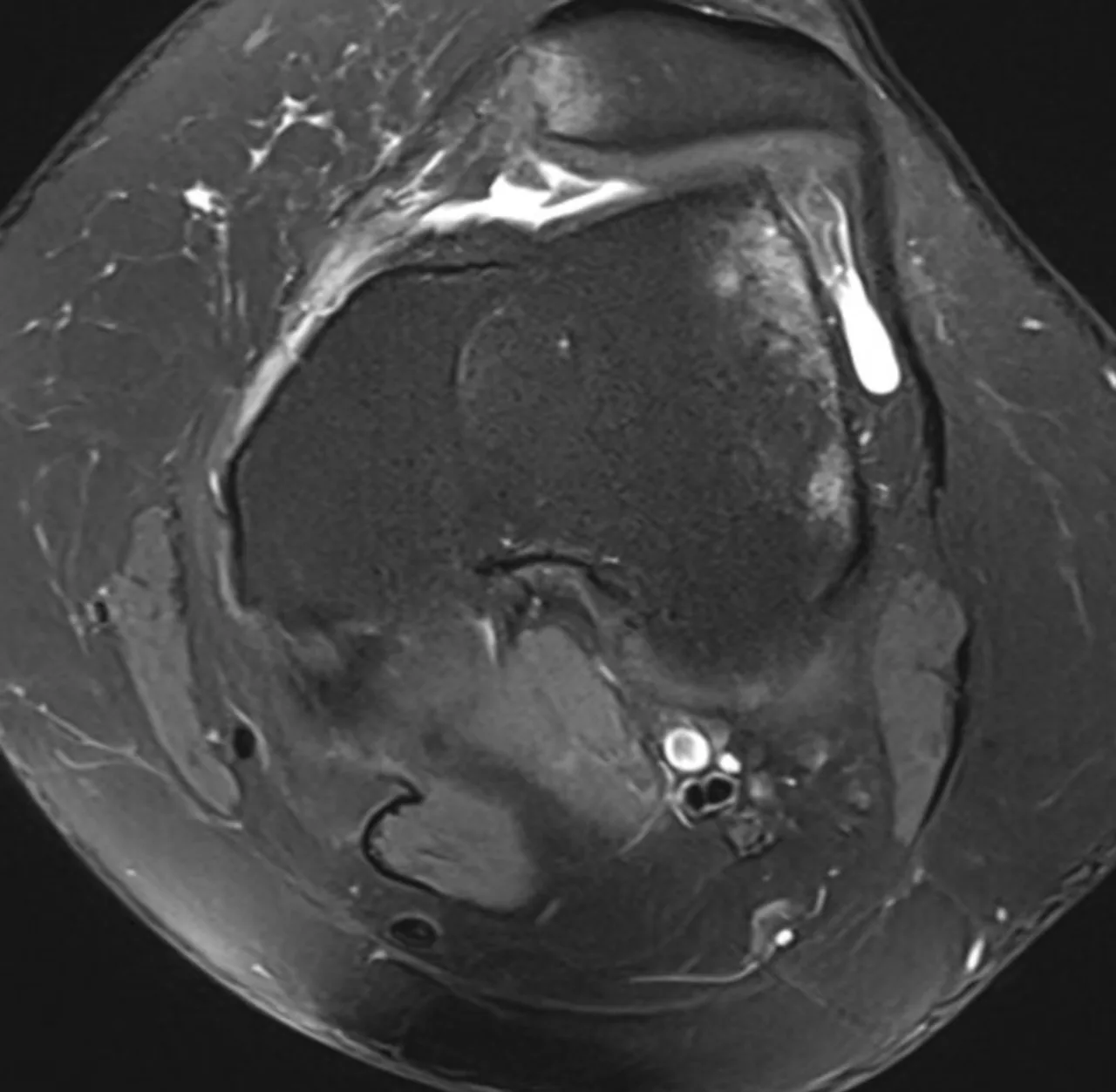
The patella normally tracks in a groove at the end of the femur. In a dislocation it slides outward, tearing the ligament that tethers it on the inner side. Some people dislocate once from a direct blow; others have anatomy — a shallow groove, a high-riding kneecap, rotational alignment — that makes it happen repeatedly.
The history usually makes the diagnosis. The exam looks for apprehension when the kneecap is pushed outward. X-rays and MRI assess the MPFL, cartilage damage on the undersurface of the kneecap, and the bony measurements that predict whether it will happen again.
After a first-time dislocation, a brace and a therapy program focused on the quadriceps, hip abductors, and landing mechanics are the standard first step, and many patients never dislocate again.
Recurrent dislocation, a loose fragment of cartilage in the joint, or anatomy that makes another episode near-certain all point toward surgery.
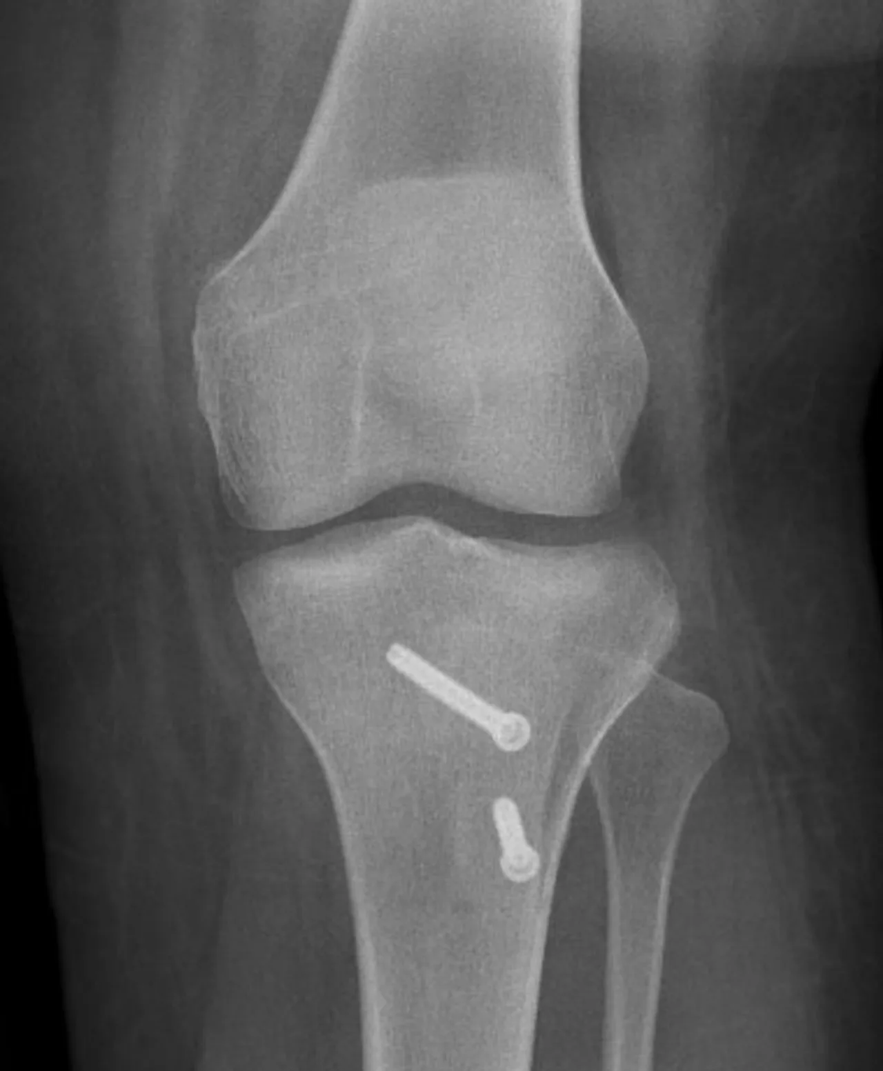
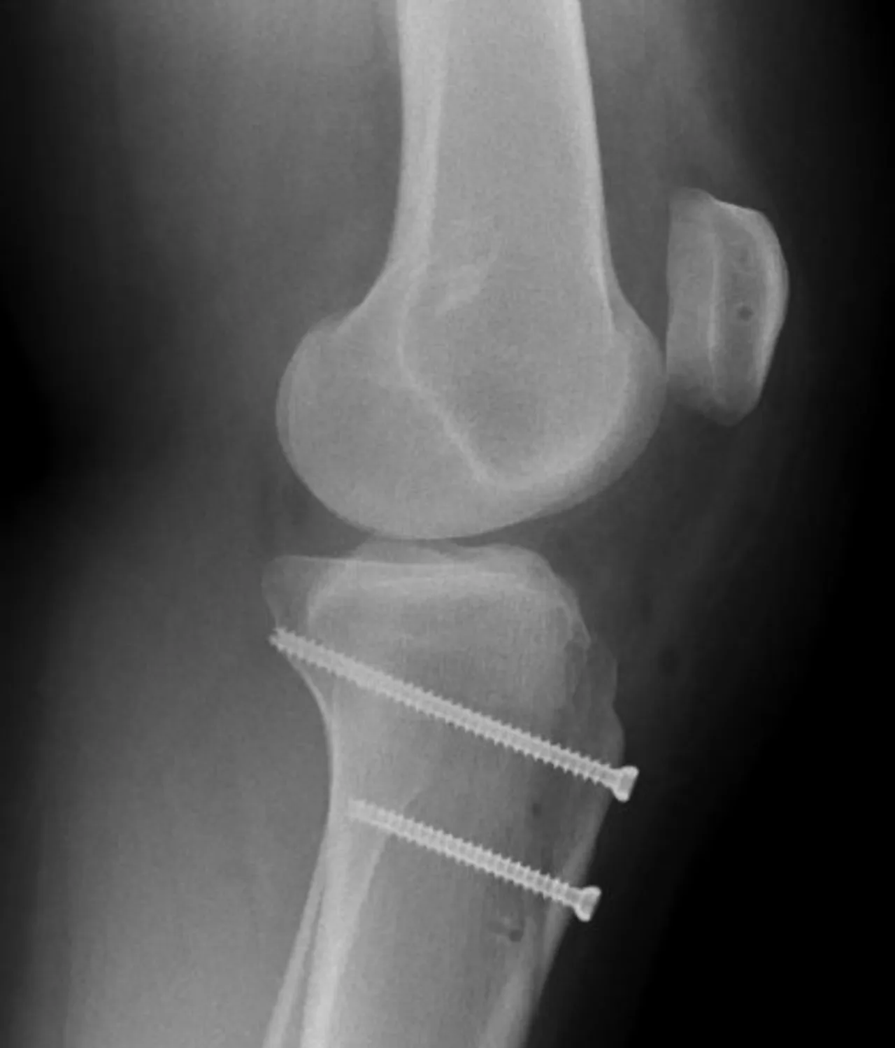
MPFL reconstruction rebuilds the ligament that holds the kneecap in place. When bony alignment is the driver, a tibial tubercle osteotomy is added to reposition the pull of the tendon. Cartilage damage is addressed at the same time.
- Is my anatomy likely to cause this again?
- Do I need bone work or just ligament reconstruction?
- What can therapy realistically fix on its own?
These pages describe how these injuries are generally evaluated and treated. They are not a diagnosis and not medical advice. Two people with the same MRI finding can need completely different plans — that is what the visit is for.