Understand your injury.
Choose a condition to read what is damaged, how it is diagnosed, what to try before surgery, and how long recovery takes. These are the conditions we see most often, not a complete list of everything we treat. If yours is not here, call the office.
Osteochondral lesions of the elbow (OCD)
Outer-elbow pain, swelling, or catching in a young thrower or gymnast — sometimes with loss of full straightening.
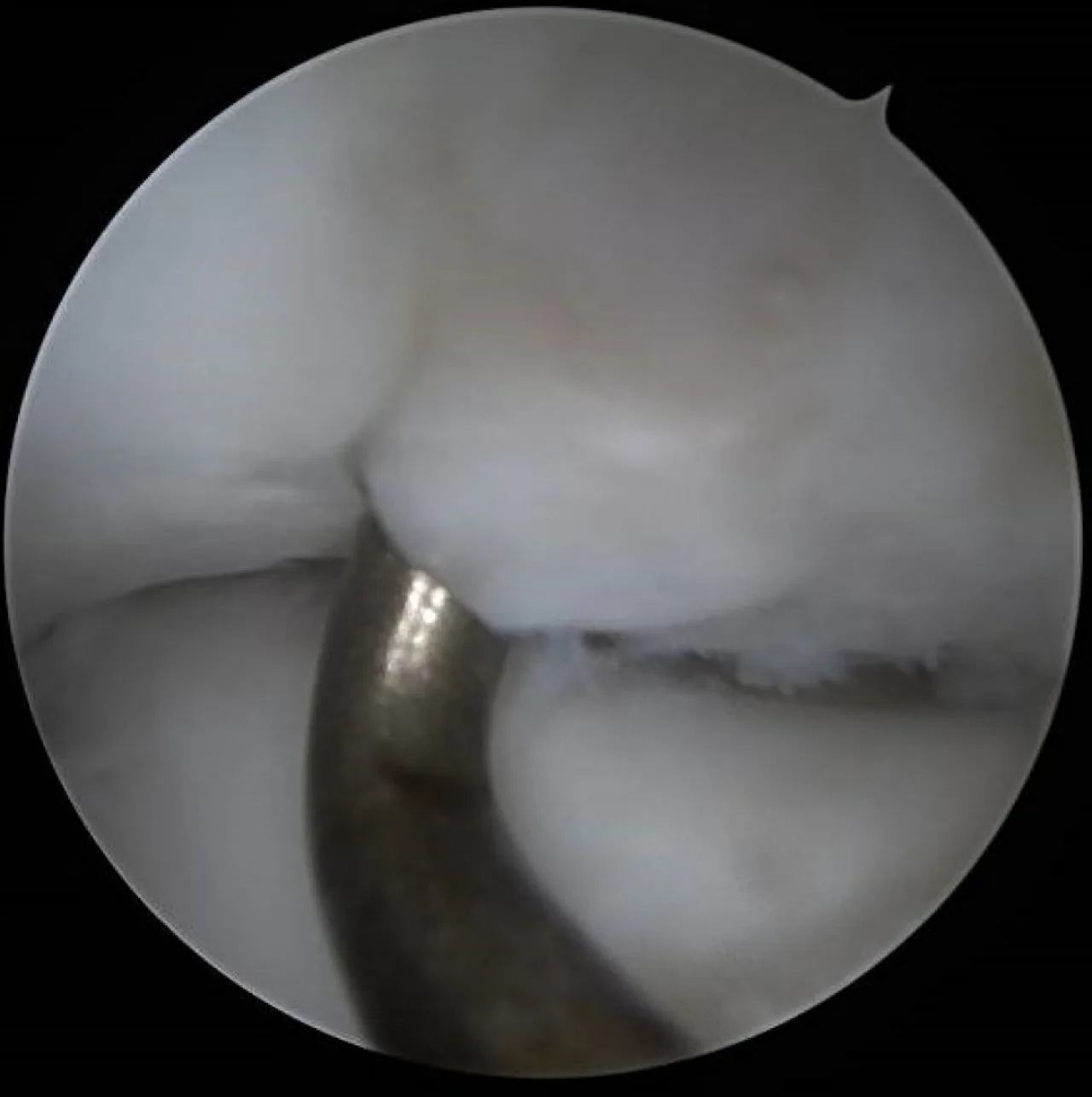
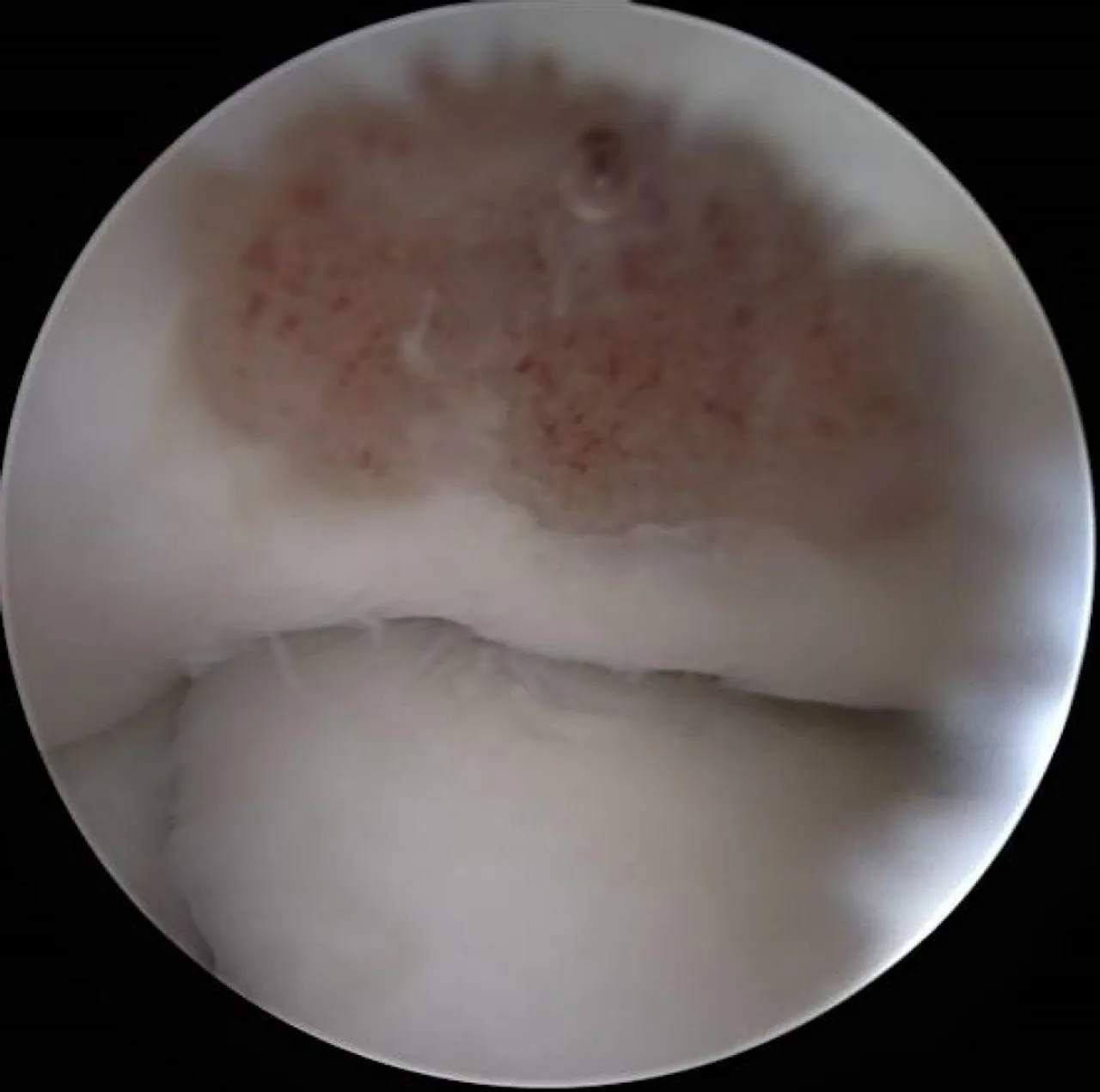
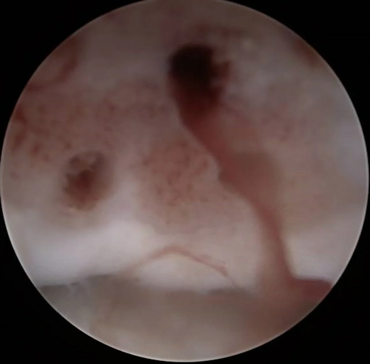
Osteochondritis dissecans (OCD) is a problem with a small area of bone and the cartilage covering it, most often on the capitellum — the rounded end of the upper arm bone on the outer side of the elbow. Repetitive compression from throwing, gymnastics, or weight-bearing on the arms disrupts the blood supply to that spot, and the bone beneath the cartilage softens. Early on, the cartilage stays intact; if it progresses, the fragment can crack, loosen, or break free as a loose body in the joint. It is seen mainly in adolescents whose bones are still growing.
Exam checks for tenderness on the outer elbow, swelling, and whether the elbow straightens fully. X-rays can show the lesion but may miss early stages. MRI is the key test — it shows whether the cartilage is intact and whether the fragment is stable, which is what determines treatment.
Stable lesions, particularly in younger patients with open growth plates, often heal with rest from throwing or weight-bearing on the arms for several months, followed by a gradual, supervised return. Follow-up imaging confirms healing before return to sport.
Considered when the fragment is unstable or loose, when a stable lesion does not heal with a full course of rest, or when loose bodies cause catching or locking.
Most procedures are arthroscopic. Depending on the lesion, that can mean removing loose fragments and stimulating healing of the base (microfracture or drilling), fixing a large fragment back in place, or — for larger defects — an osteochondral autograft transfer, moving a small plug of healthy bone and cartilage from the knee to resurface the elbow. The aim is to restore a smooth joint surface and return the athlete to their sport.
- Is my lesion stable or unstable on MRI?
- Can it heal with rest, and for how long?
- What does return to throwing or gymnastics look like afterward?
These pages describe how these injuries are generally evaluated and treated. They are not a diagnosis and not medical advice. Two people with the same MRI finding can need completely different plans — that is what the visit is for.